Herbarium (to be more exact - fungarium) of the future - Fungi


Herbarium (to be more exact - fungarium) of the future - Fungi |
|
| In this section descriptions of various live organisms (animals, plants, fungi, etc.), which could live on the Earth in Neocene epoch. The section will be supplemented as new ideas about possible ways of evolution of life will appear. If readers will not find here any species placed here earlier, it means that it is a reason to search for a new chapter in English version of "The Neocene Project". | |
|
Fungi |
Fungi - oomycetes
|
Predatory
water mold (Xenosaprolegnia carnivora)
Order: Saprolegniales (Saprolegniales)
Family: Saprolegniaceae (Saprolegniaceae)
Habitat: freshwater reservoirs of temperate, subtropical and tropical latitudes
of Old and New World, Meganesia.
Picture by Biolog
Among mushrooms there is a big group which representatives
live exclusively in water. These ones are primitive oomycetes fungi which because
of their appearance had received the common name “water mold”. Mycelium of these
fungi forms cotton wool-like white cover on rotten substrata of animal origin.
Frequently oomycetes develop on open wounds of fishes, corroding their tissues
up to bones (fungus Saprolegnia leads such way of life).
One of Neocene species of oomycetes had passed to more effective way of feeding
– it had turned to passive predator. Predatory species usually meet among soil
fungi. They form sticky heads and trapping loops, in which microscopic invertebrates
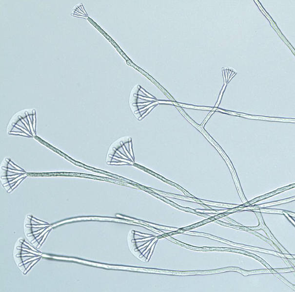
get. Predatory water mold catches prey exclusively with the help of glue. This
fungus forms special plentifully branching hyphae. Cells forming these hyphae
have elastic walls and can stretch strongly. On tips of such hyphae traps form
– they look as wide sticky heads. Each head represents a drop of glue of gel-like
consistence, “stretched” on brush-like branching terminal cells of hypha.
This oomycete develops exclusively due to live organisms caught by it. Predatory
water mold forms sticky locks about 10 cm long in still places of ponds and
rivers, among vegetation. With the help of sticky heads this fungus catches
tiny organisms like protozoans, rotifers and other worms. When small animal
is pasted to trapping head of fungus, substances from covers of its body diffuse
into slime. Their occurrence serves as stimulus for growth of cells on which
the drop of glue is stretched. If caught animal starts to try to escape, fungus
hyphae do not come unstuck from its body due to the extensibility of cellular
walls. Moreover, returning to initial position, hyphae of fungus draw caught
prey back, and it may casually paste to some other sticky heads. The cells forming
trapping head, reach a surface of prey body in some minutes. They start to emit
the enzymes destroying cell environments of prey, and sprout in its body similarly
to roots, dissolving and absorbing substances of prey body. In some hours no
rests remain from soft-bodied, and from small larvae of crustaceans and insects
only chitinous armours keep. Large colonies of predatory water mold represent
real danger to tadpoles, fry of small fishes and larvae of shrimps. But this
fungus itself serves as food to some water animals – to adult crustaceans and
molluscs. It is eaten also by some fishes and ducks.
Spores of predatory water mold are covered with protective mucous membrane,
and are easily carrying to other reservoirs on legs of waterfowl. It explains
practically global settling of this fungus, which is settled with the help of
migrating birds. Besides populations of this fungus are rather homogeneous genetically,
and it does not have any geographical variations.
In the field of an area with seasonal climate the mycelium of predatory water
mould, as a rule, dies off at formation of ice on surface of water, and survives
in winter as spores. In areas where the temperature of water does not fall below
+4 ?С, predatory water mould successfully winters as a mycelium. Spores of this
fungus sprout on the organic substratum of animal origin - usually on body of
dead small animal, on the rests of prey of any predator, or on shells of eggs
of fishes or amphibians. Proteins of substratum serve for initial development
of small lock of mycelium with several trapping heads. The further development
depends on success of hunting of this fungus.
Fungi - ascomycetes |
“Cadaver
flower” (Cadaveroflos dimorphus)
Order: Hypocreales (Hypocreales)
Family: Ergot fungi (Clavicipitaceae)
Habitat: tropics of Old World – Central Africa, Zinj Land, Southern and Southeast
Asia.
Evolution of fungi is, probably, not as appreciable, as evolution of animals
and plants. However, fungi are the major participants of processes of decomposition.
Among them there are also numerous symbiotes and parasites of plants and animals,
and some fungi have adapted to lead predatory way of life. Basically these are
microscopic ground fungi which catch various microscopic worms and other invertebrates
by loops and gyphae nets. However, among predating fungi there are also larger
species.
The territory of tropical rainforests of Old World is inhabited by large carrion-eating
dipteran species, infectiofly
(Dolichomusca infectans). It searches for carrion, using the keen sense
of smell, and lays larvae in it. Though this insect has fine sight sense, in
searches of carrion it is guided only by sense of smell, and it is actually
indifferent for it, how the substratum having a “correct” smell looks.
This feature of behaviour of insect is used by some species of fungi which breed
and live due to scavenging insects. One of these fungi is freakish “cadaver
flower”. It is a fungus rather precisely imitating carrion for attraction of
insects eating it, including infectiofly. Life cycle of “cadaver flower” includes
two phases of development. Imitation of carrion takes place at “vegetative”
stage of life cycle. From spore of fungus the mycelium grows, which develops
in vegetative rests – it is a rarity in the order to which mainly parasitic
species belong. Developing mycelium forms without fertilization a kind of “false
fruiting body”, laying directly on substratum – it is a product of evolution
of sclerotium, which now represents original “vegetative” generation in life
cycle of the fungus. This formation is vaguely similar to a flower with the
irregular-shaped petals, laying directly on the surface of substratum. Edges
of this formation are slightly raised above a substratum. Actually, “false fruiting
body” represents an apothecium characteristic for ascomycetes. This formation
is two-layer. Its surface inverted to substratum is grey and leathery, and its
middle is penetrated with numerous strings of mycelium directed into substratum.
The surface inverted upwards has jelly-like consistence, is colored reddish-pink
and has an expressed smell of decomposed meat. Spore-bearing sacs do not develop
in it, but the jelly-like mass is plentifully penetrated with threads of mycelium.
The insects involved with smell of carrion, gather on “cadaver flower” in thousands.
They creep on jelly-like mass, taste it and even leave on it larvae –this fungus
imitates a smell of decomposed meat so precisely. But then they fly out, carrying
away on legs a part of mycelium. And then, of course, having bad memory, insects
catch in the same trap again, visiting other specimens of “cadaver flower”.
As a result of flights from one fungus to another on legs of insects in the
rests of jelly-like mass parts of mycelium of genetically different specimens
of fungus. It is a stimulus for the further development of “cadaver flower”
and its passing to the second, sexual phase of development. Conjugating mycelium
of different individuals starts forming of fruiting body. Hyphae of fruiting
body penetrate into cuticle on leg segments of insect and gradually penetrate
its body entirely. The fungus infects an insect, develops in its body and within
several hours simply kills its prey. Then fungus starts to develop as saprotroph,
and the body of dead insect serves as a substratum for initial stages of development.
Spore-bearing stage of fungus considerably differs in shape from “false fruiting
body”. From stigmas, mouth and anus of dead insect true fruiting bodies emerge,
rise on thin stipes and produce spores. They represent bell-like conic heads
2 - 3 mm in diameter and up to 5 mm in height on very long stipes. The basis
of stipe is dense, but the top part, right under the head, is very flexible.
At the slightest breeze from the shaken head threadlike spores laying in sacs
among friable mass of mycelium strings are thrown out. On suitable substratum
from them the mycelium develops, forming “false fruiting body”, imitating carrion.
The idea about existence of this species of fungi was proposed by Arthropod, the forum member.
Honey-bearing
xenocordiceps (Xenocordiceps meliferus)
Order: Hypocreales (Hypocreales)
Family: Ergot fungi (Clavicipitaceae)
Habitat: northern hemisphere, temperate and subtropical latitudes, forests.
Some ergot fungi known in human epoch were parasites of insects. Sprouting in
a body of an insect, fungus gradually killed it and then formed a sporocarp,
and its spores attainted new insects. In Neocene epoch among ergot fungi new
species of such parasites have evolved. One of them is honey-bearing xenocordiceps,
which passes only a part of life cycle on insects.
Sporocarps of honey-bearing xenocordiceps develop on dead vertebrates of small
size – birds or mammals. This mushroom frequently settles on prey of various
predators and takes part in decomposition processes. Congestions of sporocarps
of this species look like thickets of grass of white color, where each “blade”
represents a separate sporocarp looking like a mouse-tail. The fungus attracts
numerous flying insects with transparent sweet drops, which are secreted on
the surface of sporocarps. But in such entertainment a real danger of death
to an insect is hidden – this liquid contains a plenty of fungus spores, which
wait while the insect will drink this liquid.
Spores of honey-bearing xenocordiceps infect an insect and sprout in its body.
The fungus keeps an insect alive within several days – it is necessary for successful
continuation of life cycle. The behaviour of the infected insect changes – now
it does not eat and does not copulate, but only searches for places where it
is better for the fungus to grow. Infected insects fly on corpses of small animals,
orientating by smell. Thus on corpses even infected butterflies may be met,
which usually are not attracted by carrion.
When a suitable substratum is found, the insect lands on it and does not move
any more. The fungus begins to grow actively and kills an insect within approximately
one hour; then it sprouts from natural orifices of its body. From died insect
mycelium strings expand and penetrate into the substratum. Substances produced
by this fungus have expressed antimicrobic properties and can kill even insect
larvae. If the fungus infects a dead animal in which maggots already live, it
suppresses their life activities: they do not eat and do not grow, perishing
gradually. On large corpses around of the fungus colony a kind of “sanitary
zone” is formed, where insect larvae do not live. The fungus gradually expands
and forms sporocarps. They have no antibiotic properties, therefore involve
new insects.
Mycelium of honey-bearing xenocordiceps mummifies tissues on which it grows.
This fungus winters in such tissues, and forms small sporocarps in the spring,
which are enough to involve new insects and to begin new life cycle.
Madness
fungus (Theriodementor murinus)
Order: Hypocreales (Hypocreales)
Family: Ergot fungi (Clavicipitaceae)
Habitat: Eurasia, except for Far North and mountain areas of Central Asia.
Picture by Biolog
Due to biologically active substances produced by mycelium
ergot fungi were used by people for medical purposes. In Neocene these fungi
began to use the rich set of chemical substances for other purposes – fungi
capable to operate the behaviour of vertebrates had evolved. One of their species
is a small parasitic fungus which has got the name madness fungus for its properties.
This species is a distant relative of honey-bearing xenocordiceps (Xenocordiceps
meliferus), which has developed the life cycle of another kind.
Madness fungus parasitizes mainly small mammals – rodents, insectivores and
less often chiropterans. This species attaints tissues, keeping its victim alive
for rather long time – for about several weeks. It changes behaviour of infected
animals considerably, making them less cautious. The infected animals begin
to behave safely and aggressively: they do not hide, run in the forest openly
and noisily, search and bite relatives and animals of similar size. Especially
dangerous among them are infected bats, which begin to fly in the daytime and
can attack even large animals. The most part of such animals perishes in some
weeks from exhaustion as they feed much worse. But more often they fall prey
of various predators. In saliva of infected animals amoeboid cells of fungus
with thin walls are found, which begin to divide actively in blood. Large animals
cope with the infection, only at the weakened individuals signs of infection
are visible: predators begin to bite their own paws or tails from time to time,
and herbivores butt or kick bushes and tree trunks. If immunity is strong enough,
the organism of an animal overpowers an infection.
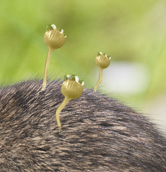
Small animals usually perish after infection. On the dead animals sporocarps
of this fungus develop – yellowish club-like heads on thin stalks. Their surface
is covered with drops of sweet liquid attracting various insects. The main carriers
of this fungus are mosquitoes. Mosquito males suck a liquid secreted on sporocarps,
and in their organisms, the fungus begins to develop. It attaints their male
gonads, and at the copulation with females males infect them. After the copulation
in female’s organism the development of the fungus begins, and it infects salivary
glands of an insect. Such females begin to attack mammals and spread amoeboid
cells of this fungus, infecting the stung animals. The birds stung by infected
insects do not fall ill – the high body temperature interferes with the development
of fungus.
Sporocarps of the madness fungus do not endure dryness and the elevated temperature,
therefore deserts of North Africa and Arabia serve as a reliable barrier to
settling of this mushroom to Africa. Also the mushroom is not cold-resistant
and consequently can not grow in areas with long winter. Bats have an opportunity
to heal from this fungus, running in the hibernation - at this time their body
temperature decreases, and the development of the fungus is slowed down a lot.
In areas of a seasonal climate the madness fungus winters in rodents, being
transferred through their bites.
Gri-gri
fungus (Grigrimycos magicus)
Order: Hypocreales (Hypocreales)
Family: Ergot fungi (Clavicipitaceae)
Habitat: tropics of Southern Asia, a symbiote of bokor ants.
Picture by Biolog
In human epoch among ergot fungi many forms parasitizing on
insects were known. In Neocene their diversity is as great, as in human epoch.
Usually such mushrooms, when infecting an insect, quickly bringing it to death
and form a sporocarp on its corpse. But one species of these mushrooms has developed
symbiotic relations with bokor ants
(Mycophoros bokor) living in forests. This species of insects has the special
caste supplied with mycangium pits on their bodies, where pieces of mycelium
of this fungus are kept. Carriers of the fungus penetrate into colonies of other
ant species and leave there mycelium of gri-gri fungus, dooming these colonies
to unenviable existence and using them as a source of food.
The fungus penetrates into organisms of insects through tracheas or through
elastic cuticle in places of joints of an armour. It begins to expand in the
host’s body, penetrating into its head and infecting its brain. As against the
majority of related kinds of parasitic fungi, gri-gri fungus does not cause
total death of the infected insects, developing in their bodies for a long time.
It changes considerably the behaviour of hosts, though does not interfere with
normal existence of their colony. All changes in behaviour become obvious at
an attack of bokor ants: the infected insects do not attack these robbers and
allow them to plunder their colony, carrying away larvae and pupae without any
obstacles. The fungus also shortens life expectancy of the infected hosts, making
it approximately one third shorter. The dying insect searches for a colony of
bokor ant by smell and “capitulates” to them, perishing on the tracks laid by
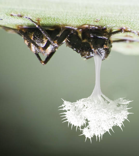
ergates of this species. The infected insect perishes, hanging back down on
the grass leaf. From a corpse of an insect a sporocarp sprouts, looking like
little thin snow-white tree with needle-like branches. Fungus carriers from
among bokor ants search for such fungi by smell and scrape off mycelium and
sporocarps from corpses of insects, hiding it in their mycangiums. The fungus
gets an advantage due to effective “address settling” of it by ants. If there
are no bokor ants nearby, gri-gri fungus can settle independently with the help
of spores and infect new hosts, not involving bokor ants.
The idea about existence of this fungus species was proposed by Morgot, the forum member.
Tempting
inannomyces (Inannomyces hypervenustus)
Order: Hypocreales (Hypocreales)
Family: Cordycipitaceae (Cordycipitaceae)
Habitat: Eurasia, forests of temperate and subtropical zone.
Parasites represent an integral part of ecosystems. Every biological species
has a set of the specific and nonspecific parasites adapted to the life on the
present species. Evolution of parasites takes into account features of the host’s
physiology, and the parasite fits its own life cycle to them for more successful
spreading.
Butterflies in temperate and subtropic zones of Eurasia are parazitised by the
original fungus – tempting inannomyces, which is a close relative of fungi of
Cordyceps genus. It develops in bodies of insects, killing them, but in order
to breeding, it uses a natural appetence of heterosexual insects to each other
during their breeding. This fungus species is spreaded in a population of insects,
increasing the production of pheromones at the infected adult individuals ready
to breeding, and infects insects at their copulation. Hence the name: Inanna
is a goddess of fertility and love in Mesopotamia.
Influence of this fungus to organisms of butterfly male and female is different.
Inannomyces is capable to exist in male organism for a long time, not killing
it. At butterfly males the fungus affects only genitals, keeping an insect alive
during rather long time: it is necessary for distribution of the species, because
the fungus is transferred to females at the copulation. In fact, it is an original
“venereal disease” of insects, inevitably resulting in lethal outcome. In male’s
genitals, fungus forms tufts of filaments, which easily crumble to separate
fragments and stick to the surface of genitals. Getting at the moment of copulation
to the female’s organism, fungus quickly expands in its reproductive system
and causes reorganization of its physiology. Eggs in female’s organism stop
their development – they turn to a substratum, on which the further development
of this fungus goes on. Female stays alive at this time, however it becomes
sterile and starts pheromone secretion actively, involving as many males as
possible, and they catch this fungus. Later the fungus sprouts in female’s tissues,
causing fast degradation of its organism. Dying female searches actively for
a fodder plant of its species, lands on it and quickly perishes. From its organism
through its mouth, spiracles and an aperture of hindgut sporocarps of the fungus
grow out – threadlike formations up to 30-35 mm long of brown color with white
spherical heads spreading spores. Spores spread with the wind, and a part of
them with a high degree of probability can infect the caterpillars of the butterfly
fed on this plant.
Within one summer this fungus can give some generations, depending on the duration
of life cycle of the host species. In conditions of a seasonal climate, the
fungus winters in insects at the stage of pupa, from which the infected sterile
insects burst, having normal sexual behaviour, reacting to pheromones and infecting
healthy individuals with this fungus.
Aphid
brush fungus (Aphidinervum sterlilsator)
Order: Hypocreales (Hypocreales)
Family: Cordycipitaceae (Cordycipitaceae)
Habitat: Eurasia, temperate latitudes, the parasite of plant lice.
Parasitic fungi affecting various species of insects were widely known in human
epoch. The variety of insects in human epoch has suffered relatively small damage,
and quickly compensated it in during the ecosystem restoration period. In Neocene
the diversity and availability of insects as a food source for fungi grew in
parallel to restoration of natural communities and distribution of forests.
At this time in nature the new species of parasitic fungi have evolved, having
mastered the development on various kinds of insects.
Aphids (plant lice) have suffered rather small damage in human epoch, and in
Neocene they still represent a characteristic component of entomofauna of temperate
latitudes, having numerous symbiotes and parasites. One parasite of plant lice
is a tiny fungus named as aphid brush fungus. The sporocarp of this species
is reduced in great degree because of the tiny size of its host species, and
resembles a tiny translucent brush with thin stalk no more than 6 mm long. Inside
a bunch of hyphae on the tip of a sporocarp ascs with wind-dispersing spores
form. Spores of this fungus are also distributed on the body surfaces of ants
and insects eating plant lice – they both are frequently found in colonies of
plant lice and promote the infection of new colonies.
The first stage of infection of a plant louse takes place on the surface of
its body soiled with sugary excretions (honeydew), where the spores sprout.
Hyphae of the fungus penetrate into the body of insect through spiracles or
gland ducts, and the fungus begins to develop inside the body of plant louse,
transforming it to a kind of zombie capable only to eat and actually not reacting
to external stimuli like touches of an ant gathering honeydew. The most part
of hyphae of fungus sprouts to ovoducts of an insect and destroys posterity
formed there. The fungus keeps a plant louse alive for a long time, and uses
the nutrients synthesized in its organism, interfering simultaneously with its
breeding. It actually turns to the supplier of food for a fungus. If it is necessary,
the infected insect can move, and even drive away its congeners from the most
attractive fodder sites.
When the fungus is ready to breeding, from plant louse’s ovoducts sporocarps
of the fungus grow like tiny brushes. The infected insect may be easily distinguished
among healthy individuals by a long thin brush of sporocarp of the fungus sticking
out of its body upwards, and by less transparent body, if body covers at the
present species are normally translucent – because of gyphae of the fungus inside
the insect’s body. Spores are easily carried away by slight streams of air in
underbrush and infect plant lice from other colonies, but the more reliable
way of their distribution is an insect eating plant lice.
In an autumn, when in colonies of plant lice wintering individuals begin to
develop, activity of the fungus decreases. It still infects insects, but enables
them to breed. Such individuals lay wintering eggs already infected with a fungus.
In the spring from them nymphs burst, in which the development of this fungus
goes on. Also this fungus transforms some slightly infected females to wintering
ones, giving them an opportunity to winter and to continue to feeding in the
spring to enable the fungus to disseminate its spores. Also aphid brush fungus
winters as spores on bodies of predatory insects.
“Forest
goblet” (Eupelicomyces multicapitatus)
Order: Pezizales (Pezizales)
Family: Pezizaceae (Pezizaceae)
Habitat: Chile, humid Nothofagus forests.
Moderate – cold areas of Southern hemisphere in Neocene had kept approximately
the same area, as in human epoch. Though South America had moved a little aside
the South Pole, climate warming in Neocene has caused displacement of border
of warm-moderate climatic zone to poles. Therefore the climate more-less similar
to climate of the most part of Eurasia, in Southern hemisphere was generated
only at the far south of South America. But also here it undergoes great influence
from the part of ocean. Therefore moderately cold snow winter is characteristic
for this area. Summer at the far south of South America is humid, about rather
small amount of dry and hot days. Such weather is favorable for growth of various
mushrooms.
Forests at the south of South America are formed by various species of southern
beech (Nothofagus) – the kind of plants growing in temperate areas of Southern
hemisphere from Mesozoic. Forests of this plant form a habitat for plenty of
mushroom kinds. Also it is very remarkable, that right here instead of rich
in life forests of Amazonia, the largest mushroom of South America, “forest
goblet”, grows.
The body of mushroom consists of false tissue – plectenchyma, which represents
a texture of mycelium fibers. It is lack of strenghtening tissue, therefore
it is absolutely not feasible for a mushroom problem to grow up to the size
of tree. “Forest goblet” exists in fact at the edge of mechanical opportunities
of plectenchyma. It is the huge mushroom reaching one meter height. High humidity
of air favours to growth of its huge fruit body. The fruit body of “forest goblet”
represents huge ramified apothecium of yellow color with brownish shade. Edges
of “pileus” have darker color – they are orange-brown. A consistence of fruit
body is elastic, and the bottom part of stem of mushroom is gristly. Its “pileus”
has strongly concave form, and after morning fog a lot of water accumulates
in it. Streaming down along internal surface of apothecium, water washes off
spores of mushroom. Inside the mushroom small birds frequently arrange original
“swimming baths” for themselves, after which they carry spores of mushroom on
their plumage. In middle part the stem of fruit body branches and forms some
small cup-like slightly irregular-shaped pilei. If branching of stem takes place
higher, instead of one “pileus” of rounded outlines “Siamese twins” – two, three
and even more “pilei” grown together are formed. “Forest goblet” is not poisonous,
but has bitter taste protecting this mushroom from rodents and birds.
There is one more secret of large size of “forest goblet” – this mushroom forms
mycorhiza with southern beech, receiving from it some of organic substances
instead of mineral substances. The mycelium of “forest goblet” actively participates
in decomposition of wood litter, and its fruit bodies are formed, when in forest
its greatest amount gathers – closer to middle dawn (in Southern hemisphere
approximately in the beginning of April). “Forest goblet” forms mycorhiza with
large species of Nothofagus forming a basis of forests of temperate zone of
Southern hemisphere.
The close species also lives in forests: crested
mushroom (Eupelicomyces cristatus). It is a mushroom of considerably
smaller size, than “forest goblet”: the maximal height of fruit body is no larger
than 15 centimeters. But its fruit bodies reach the significant size, because
“pilei” of this mushroom are not rounded, but merged to longitudinal crests.
The length of such fruit body may reach 30 centimeters. At the fruit body of
crested mushroom some crests with spore-producing surface develop. Their edges
are dissected to rounded lobes of reddish-brown color with bright orange edges.
This species does not form mycorhiza and lives as saprophyte. Its fruit body
emits the unpleasant smell involving insects, and more often spores of this
mushroom are distributed by beetles. Fruit bodies of crested mushroom appear
in forest at the end of summer and an early autumn.
Purgative
mushroom (Dulcipeziza purgans)
Order: Pezizales (Pezizales)
Family: Pezizaceae (Pezizaceae)
Habitat: subtropical and tropical forests of Eastern and South-Eastern Asia,
some locations in Hindustan; wood litter.

Picture by Biolog
A variety of life strategies related to interaction with animals
at various stages of life cycle was typical for fungi of human epoch. In Neocene
evolution of mutual relations between fungi and animals has continued, and some
fungi have developed original strategies of survival.
On wet wood litter in forests of Eastern and South-Eastern Asia purgative mushroom
lives. It is a representative of ascomycetes, which in due course of evolution
has developed very simple strategy of mutual relations with the animals living
in the neighbourhood. This mushroom is saprophyte on leaf litter; its mycelium
penetrates it at the depth of several centimeters under the surface, where sufficient
humidity for its growth is always kept. Seasonal prevalence in formation of
sporocarps in the southern part of the range is not expressed; at the north
of the range sporocarps of this mushroom form mainly at the end of spring and
in the beginning of an autumn, when there is enough amount of rains. In the
summer at the north of the range sporocarps are formed only at sufficient humidifying
of the ground.
Sporocarp of purgative mushroom represents large bowl-like apothecium up to
half meter in diameter, of dense, gristle-like consistence. Its edges are raised
on 3-4 cm above the ground. From outside on the sporocarp grey аудедшлу cover
passing to mycelium is present. Apothecium is full of dense slimy mass containing
spores and has very bright citreous colouring of internal part. Contents of
apothecium swells during the rain and rises above its edges. This mass has pleasant
taste, and large herbivorous mammals willingly lick it off. Bright colouring
makes this mushroom better visible in twilight of underbrush. The mass with
spores, however, has laxative effect – hence the name of this mushroom. This
is an adaptation for settling: having eaten it, the animal has time to walk
off far enough. Due to activity of intestines of an animal, spores of mushroom
pass through it safely and drop outside in liquid manure. Manure acts as a nutrient
medium for development of spores. In addition, when licking off spores from
several mushrooms, animal promotes the occurrence at the small site of forest
of myceliums of several unrelated caryotypes at once; it favours to formation
of new sporocarps of mushroom.
Also this mushroom is settled on feet of birds, which perch on edges of sporocarp
and peck off the spore-bearing mass. It, probably, explains the existence of
several sites of purgative mushroom growth in Hindustan, where the related species
– vomitive
mushroom (Dulcipeziza vomica) – lives. This species is similar to purgative
mushroom, but differs from it in appearance and properties. Its sporocarps reach
only 30 cm in diameter, and have lighter colouring of their internal parts.
The swallowing of slimy mass with spores causes vomitting in the majority of
herbivorous mammals, but rodents and primates eat it without any bad consequences.
Carnivores frequently use this mushroom for stimulation of regurgitation of
swallowed wool.
“Frog’s
cup” (Dendrocalyx raninus)
Order: Pezizales (Pezizales)
Family: Pezizaceae (Pezizaceae)
Habitat: forests of Equatorial Africa, forest canopy.
In due course of evolution various species of live organisms belonging to systematic
groups unrelated to each other can develop mutually advantageous strategy of
cooperation – they enter symbiosis. On tall trees of tropical forests of Africa
the unusual union of arboreal deafening
frog (Decibellator abalienarus) and a mushroom called “frog’s cup” was developed.
Like the majority of the related forms, “frog’s cup” mushroom is saprophyte.
Its mycelium sprouts in cracks of tree bark and in forked branches, where epiphytic
plants grow and the foliage and other organic dust accumulate. “Frog’ cup” frequently
grows among thickets of epiphytic ferns or orchids, actively taking part in
decomposition of organic matter and forming the mycorhiza with epiphytic plants.
Sporocarps of “frog’s cup” are rather large – these are cuplike apothecia with
high edges, in which up to 300 milliliters of rain water accumulates. Apothecium
has a smooth surface and rusty-brown color. Edges of sporocarp are slightly
wavy, and an external surface is rough to the touch. Sporocarps of various ages
grow in groups of 3-4 ones. Duration of existence of each sporocarp is about
3 weeks.
Internal surface of sporocarp of “frog’s cup” is smooth. On it ascs with spores
are located and a jelly-like mass is secreted – it is the recipe strengthening
the union of mushroom and frog. The deafening frog breeds exclusively in water
accumulated in sporocarps of this mushroom. During their development, tadpoles
scrape the mass secreting by “frog’s cup” mushroom and eat it. In this mass
there is a lot of spores of this mushroom. Having got in a digestive path of
the tadpole, many spores perish, but some sprout and infect a growing tadpole.
It easily endures infecting by mushroom and does not decrease rates of growth.
The mushroom is located mainly in epithelium of hindgut and is capable to exist
for a long time in an organism of this amphibian. When tadpoles pass metamorphosis,
they carry mycelium of “frog’s cup” to other trees. In the first weeks after
metamorphosis the young frog distributes mycelium with its feces. Organism of
animal gradually fights off the infection and is cleared of mushroom, but during
the time of infection frog manages to carry mycelium far enough from the place
of its birth. Frog’s feces serve as a substratum for initial development of
mycelium.
Spores of “frog’s cup” also settle on feet of birds using sporocarps of mushroom
for bathing or water drinking.
Spring
ice mushroom (Cryomorchella vernalis)
Order: Pezizales (Pezizales)
Family: Morels (Morchellaceae)
Habitat: steppes of Three-Rivers-Land, Southern Ural, steppes of Southern
Siberia.

Picture by Alexander Smyslov
(background: herd of porcippulas)
Mushrooms are very important component of ecosystems. These
organisms occupy prevailing position among saprotrophs, and due to their vital
activity processes of decomposition of dead organic substances proceeds much
faster.
In steppes of Three-Rivers-Land each summer grass plentifully grows on. Herds
of herbivorous mammals feed on it, but they succeed to eat not all growth of
graminoids. When winter begins, rains and small layer of snow force the withered
grass to the ground, and it starts to decompose. In early spring on slopes of
hills warmed up by sun among yellowish-brown last year's grass mushrooms of
white color appear. It is a species of mushrooms which plays a determining role
in process of decomposition of vegetative rests in steppes of Eurasia with seasonal
climate and cold winter. For the cold endurance and time of active growth it
is named as spring ice mushroom.
This ascomycete has rather low competitiveness, and can be considered as original
analogue of ephemeral plants among mushrooms. Its fruit bodies cannot be met
in summer when in steppe other species of mushrooms grow. Their mycelium strongly
suppresses growth of mycelium of spring ice mushroom, and this species as if
vanishes from steppe to the most part of year. Within several months, since
late spring up to middle of an autumn, spring ice mushroom is kept only as resting
spores in ground.
When autumn and winter colds begin, the majority of mushrooms stop growing,
and their mycelium develops much slower. Colds serve as stimulus for sprouting
of spores of spring ice mushroom – its spores develop, when the day time temperature
falls up to +5°С, and there are frosts at night. From late autumn till early
spring the mycelium of this species develops on rests of last year's grass.
In some places the mycelium of spring ice mushroom forms extended textures under
layer of rotten grass. When the top layer of ground freezes through, growth
of mycelium stops, but after the first days of thawing weather it renews.
In the beginning of spring, when the sun warms up the top layer of ground enough,
spring ice mushroom for a short time turns to sun-worshipper. From under layer
of last year's grass its fruit bodies appear plentifully. They may be so numerous,
that some slopes from apart seem strewn with snow. The fruit body of spring
ice mushroom is separated to small wrinkled pileus similar to dried up apple
about 2 – 3 cm in diameter, and thin stem up to 15 cm high. These mushrooms
grow very quickly, and the fruit body develops completely during approximately
3 – 4 days. Despite of fragile appearance, this mushroom has excellent vitality.
It endures frosts and a snowfall which frequently come back in spring.
Spores of spring ice mushroom develop on the surface of its pileus, in thin
layer of slime. The main carriers of spores of spring ice mushroom are steppe
herbivorous mammals – porcippulas,
harelopes and other animals. After winter food
shortage herbivores willingly bite off pileuses of this mushroom, despite of
its bitterish taste, and carry spores of mushroom with their manure. The cover
of spores of spring ice mushroom is rather strong, and it is not damaging staying
in gastroenteric path neither of mammals, nor of grubs of dung beetles.
Velvet
Guano Mould (Speleomucor nycticoprophagus)
Order: Eurotiales (Eurotiales)
Family: Trichocomaceae (Trichocomaceae)
Habitat: New Zealand, humid floors of caves inhabited by roosting bats, large
growths on mounds of decaying guano.
The spread of mankind across the globe, taking with them produce and industry,
resulted in many of their afflictions and commensals spreading alongside them.
Aspergillus mould easily spread to New Zealand via spores that settled upon
produce and goods brought by man, and even afflicted the perishable goods brought
there by people.
This common mould has various descendants in the warmer, humid parts of Aotearoa,
and many other parts of the world, but the most notable form is the Velvet Guano
Mould.
This species typically grows in clusters which coat decaying guano, on the floors
of cave-roosting bat colonies. Individual growths may become so thick as to
have a raised, bumpy texture of small knobs, and its texture resembles a sickly
greenish-black velvet. As it is actively involved in the decay of bat guano,
it partly serves as a commensal and symbiote of various small bats, and as such
does not infect the respiratory systems of the bats with which it co-habits.
Its spores may infect and cause aspergillosis in animals with a lower metabolism,
such as frogs and lizards. Growths occur in areas of cave which are rich in
guano, but in which the air is not too stale, as it requires oxygen to grow.
This mould also very much likes high levels of humidity, and the warm confines
of a bat-crowded cave are ideal for it.
Spores that spread to neighbouring areas may take hold on rotting vegetable
matter or fallen fruit. This mould is asexual, and grows very prolifically when
it has taken hold. Various kinds of scavenging and mould-eating insect, such
as beetles, enjoy feeding on it.
This fungus species was discovered by Timothy Donald Morris, Adelaide, Australia.
Wasp
nest cyttaria (Cyttaria nidus-vespae)
Order: Cyttariales (Cyttariales)
Family: Cyttariaceae (Cyttariaceae)
Habitat: the south of South America, forests.
Among characteristic representatives of mycoflora of South America there are
Cyttaria mushrooms – ascomycetes of very characteristic shape parasitizing on
various species of southern beech (Nothofagus) forming forests in the area of
temperate climate at the far south of the continent. They have survived in epoch
of global ecological crisis together with their host trees, and in Neocene,
their evolution has continued. In southern beech forests of Patagonia and Tierra
del Fuego of Neocene epoch wasp nest cyttaria lives – it is a parasitic mushroom
living on plants of Patagonian
false hazel (Nothavellana antipodorum), the representative of Nothofagaceae
family. This mushroom prefers humid microclimate of a underbrush where its host
plant grows.
Sporocarp of this mushroom has wide conical shape; it ma frequently be extended
along the branch of host tree, resembling wasp comb (hence the name). Sporocarps
are formed under bark, and in due course of growth they break it off and expand;
they are usually formed on the bottom side of the branch and extend from top
to bottom while apothecia of the mushroom ripen and open. On vertical tree trunk
they grow like a chain of small sporocarps with easily recognizeable structure
of the bottom surface, being vaguely similar to sporocarps of a bracket fungus.
The length of normally developed sporocarp may be up to 20-30 cm at thickness
of about 2-3 cm. The flat bottom side bears numerous deepenings of apothecia
immersed into its pulp; at still growing sporocarp they are closed with thin
membrane breaking while it grows. An arrangement of apothecia on the surface
of sporocarp is almost ordered, resembling a pattern of cells in combs of hymenopters.
The young sporocarp at the stage of active growth is white or pale yellow; mature
sporocarp has an expressed yellow colouring, darker in depth of apothecia, with
brown or orange shade. At the ripening, sporocarp emits well distinct putrefactive
sweetish smell. Spores of this species are carried by beetles, and also are
distributed in feces of birds or tree-climbing mammals. Sporocarps growing lower
above the ground are willingly eaten by ground-dwelling herbivorous mammals,which
also disperse spores of this mushroom.
Layered
carpet fungus (Haplocarnomyces stromatus)
Order: Carnomycetes (Carnomycetes)
Family: Protocarnomycetes (Protocarnomycetidae)
Habitat: Europe, deciduous forests of southern slopes of the Alpes.
In Neocene among ascomycetes the distinct group of carnivorous mushrooms – carnomycetes
– had evolved. Their feature is formation of special trapping structure - pseudocarpophor.
As against true sporocarp, pseudocarpophor is formed from haploid mycelium and
lacks an ability to form spores. Its function is exclusively providing of a
fungus with nutrients from the caught small animals.
At the majority of carnomycetes pseudocarpophor has a sophisticated shape and
represents a trap, where insects are kept with the help of glue, strong “cotton
wool” of hyphae or internal cavities, from which the way out is difficult. However,
in Neocene nature there are also representatives of primitive group of protocarnomycetes,
at which the primitive structure of pseudocarpophor is kept. One of their species
is layered carpet fungus.
This fungus develops in humid microclimate of the deciduous forests growing
on southern slopes of the Alpes. It can be met on the bottom side of tree branches,
and on moss “cushions” and on the bottom side of stones. Actually, the pseudocarpophor
is almost not present at this species: it represents simply dense mat of hyphae
secreting sticky liquid, atop of which there is friable “cotton wool” of hyphae
with dense cellular walls. This fungus resembles externally a cottony layer
with casual dust and insectspasted to to it.
The trap for insects works very simply: the fungus develops in places, which
are frequently used as a shelter by flying insects. In addition, it involves
insects with a smell. When the insect lands on pseudocarpophor of this fungus,
its legs immediately sink in “cotton wool”. Careless movements of an insect
result to that its legs get deeper in this liquid and an insect appears pasted
even harder. Trapped insects gradually die and are envepoled with mycelium quickly
sprouting into them. Mycelium gradually covers them and forms a new layer atop
of previous one. Old multilayered mycelium is usually loose and may be torn
off by wind blows, but of the rests of hyphae on a substratum new layer is formed,
continuing to catch insectsin proper way.
Sporocarps of layered carpet fungus are formed in a place of contact of two
genetically different parts of mycelium. They represent tiny bowl-like apothecia
of white color with smooth edges, which are placed directly on a substratum.
Tailed
aphidiolysia (Aphidiolysia caudata)
Order: Carnomycetes (Carnomycetes)
Family: Protocarnomycetes (Protocarnomycetidae)
Habitat: Europe, Northern Africa, Near East.
In due course of evolution in the group of carniforous fungi carnomycetes some
families have diverged, differing from each other in a structure of trapping
sporocarp (pseudocarpophor). But along with the specialized representatives
of carnomycetes in Neocene nature there are representatives of an early clade
of radiation of these fungi, which pseudocarpophors are underdeveloped or absent
at all. These fungi, however, have developed their own effective strategy of
survival connected to predatoriness.
Tailed aphidiolysia is a tiny representative of carnomycetes: the size of its
sporocarp is measured in millimeters. This fungus eats plant lice – small and
intensively breeding insects. Due to this circumstance this fungus has virtually
unlimited source of food.
Life cycle of aphidiolysia begins, when this fungus gets on sugary honeydew
– dung of plant lice. Spore quickly sprouts and infects an insect, penetrating
into its hindgut. The fungus gradually infects all internal bodies of a plant
louse: its hyphae sprout between cells and fill in the body of an insect. The
plant louse infected with fungus feeds actively and emits pieces of mycelium
of fungus with its honeydew. If such honeydew will be eaten by ant, fungal hyphae
from its mandibles can easily infect other plant lice. The infected plant louse
changes externally: it gets “fat”, its covers are stretched hard, and the body
at species with a transparent body becomes muddy because of fungal hyphae. The
infected plant louse lives few days only. During the last hours of its life,
insect stops feeding: hyphae of fungus clog intestinal tract lumina and sprout
into its proboscis. Through the proboscis hyphae of this fungus will penetrate
into vessels of a fodder plant, where plant lice live. Inside a plant hyphae
keep viability for any time, but later perish inevitably.
However, some fragments of mycelium succeed to reach the next host: they are
soaked up by other plant lice of a colony, which also become infected. If plant
lice in a colony feed very close to each other, the fungus infects them one
by one, directly penetrating with its mycelium into their proboscii.
Sexual breeding of tailed aphidiolysia is extremely seldom, but the fungus breeds
successfully, forming sporocarps without fertilisation. From a hindgut of a
dying plant louse a sporocarp sprouts – it is a reduced apothecium, peaked threadlike
structure up to 3 mm long, at which top ascs with spores are formed. When spores
ripen, the top of a sporocarp “becomes diluted” and turns to a drop of a viscous
sweetish liquid. It involves ants, which lick off this lure, carry spores of
the fungus and infect new plant lice.
Coccid
tentacled fungus (Coccidiovora tentaculata)
Order: Carnomycetes (Carnomycetes)
Family: Protocarnomycetes (Protocarnomycetidae)
Habitat: tropics of Southern Asia.
Despite lacking complex traps, carnivorous fungi from among protocarnomycetes
appear successful invertebrate trappers. Some of them are specialized in feeding
on animals that are virtually not capable to get into traps of other carnomycetes.
Prey of such fungi includes sedentary insects – coccids (mealybugs and scale
insects). Due to their sluggishness up to complete immovability, the fungus
is capable to pursue them.
This fungus has very small size – mycelium covers an area measured in centimeters
(approximately correspondent to a colony of insects it feeds on). Sporocarp
does not exceed one centimeter in height. Pseudocarpophor (trapping sporocarp)
closely resembles mycelium in appearance and is not bordered anatomically from
it.
Infection of a coccid colony begins from germination of spore of this fungus
on sugary excretions of insects which remain on the surface of plants inhabited
by them. It is the first substratum for development of mycelium, and fungal
hyphae begin to grow in a direction of an insect attached to a plant. The chosen
victim has actually no chance to escape: hyphae of a fungus grow faster than
this insect can creep being a nymph. Adult coccids of many species completely
lose an ability to move; therefore fungus inevitably grows up to them and inserts
its hyphae into apertures of spiracles and hindgut. Fungus changes physiology
of an insect for any time, forcing it to suck plant sap more actively, but all
nutrients are used exclusively to the growth of a fungus. Secretions of fungus
force insects to molt once again, getting some nymphal features and growing
up, but at this stage the whole body of an insect is already penetrated with
mycelium of a fungus, and there are irreversible changes in it, leading later
an insect to death. Only nymphs not lost abilities to move can escape from this
fungus, but they also frequently perish, if fungal hyphae grow up to them.
At early stages of development coccid tentacled fungus does not form sporocarps.
It takes place when the fungus infects enough of insects and gets necessary
amount of nutrients. For infection of new individuals fungus forms band-like
strings consisting of mycelium which creep on the surface of tree bark, being
guided by smell of insects. These strings represent heavily modified pseudocarpophor.
In dry air pseudocarpophor can reach the insects attached at the distance of
2-3 cm, but in conditions of humid climate, using cracks in a bark of a plant
where a colony of insects is feeding, it can move to 10 sm and farther in searches
of insects.
Fungus gradually kills the infected insects and finally devastates their colony.
Breeding of this species more often takes place in asexual way; inside a body
of an insect sporocarp representing tiny (up to 2 mm in diameter) cleistothecium
on a thin stalk is formed. Sporocarp breaks through a body of an insect on its
back and grows, waiting while any beetle or wasp dispersing spores of this species
will eat it. Spores of coccid tentacled fungus are distributed also on legs
of flies and butterflies fed on coccid excretions.
Underground
trapping fungus (Glutinomyces viscosissimus)
Order: Carnomycetes (Carnomycetes)
Family: Protocarnomycetes (Protocarnomycetidae)
Habitat: Eurasia, forests and steppes of temperate climatic zone.
In due course of evolution the majority of representatives of Carnomycetes order
has adapted to feeding on small prey – insects and spiders, occasionally on
worms. But some representatives of carnomycetes are adapted to feeding on larger
prey; it is remarkable that such adaptation was developed at protocarnomycetid
fungi, the least specialized in the anatomic relation ones. One of their species
is underground trapping fungus adapted to feeding on very large prey – digging
insects and small rodents.
Mycelium of underground trapping fungus grows in thickness of the top layer
of ground. It develops especially actively in immediate proximity from the large
cavities in ground – more often right near to burrows of small-sized animals.
Possibly, the development of mycelium is stimulated by air circulation in ground.
Having reached a hole of an animal, mycelium changes its pattern of growth and
forms pseudocarpophor – hard hyphal carpet on a surface of floor and walls of
a burrow. Initially on walls cotton wool-like layer appears, which in several
night hours very quickly accrues and turns denser. On it drops of very viscous
glue appear – they are intended for keeping of prey. In holes of small diameter
fungus expands along the arch of a hole, and from there drops of glue hang down.
Such trap may wait for prey during about one week, but more often prey is trapped
much sooner. When the animal tries to cross such trap, it is pasted to sticky
carpet. Carbonic gas and organic substances emitted by prey stimulate germination
of fungal hyphae, which gradually envelop prey. As a rule, small rodent or an
insect quickly chokes, when glue clogs its respiratory system or tracheas.
Fungus produces effective antibiotics interfering the rotting of prey. Also
they prevent settling of a mould on prey and suppress the development of insect
larvae, which may become food competitors of a fungus. The digestion of a cricket-sized
insect takes about two day, and the body of a rodent is digested almost completely
within one week. Only wool, bones or chitinous armour remain not digested.
Sporocarp of underground trapping fungus develops under the ground near the
surface. It has rounded shape and is similar to a truffle. Surface of sporocarp
is covered with a thin tunica, under which spore-bearing surface covered with
deep flexuose plicae and cristae is hidden. In fact, sporocarp of this fungus
represents a deeply modified apothecium secondarily covered with a tunica from
above. Sporocarp of underground trapping fungus emits expressed “mushroomy”
smell involving beetles that carry its spores. Sporocarps of this species may
be eaten frequently by large herbivorous mammals. Spores in many cases pass
through a gastroenteric path of an animal, keeping their viability, and later
sprout in manure of an animal.
Parasitic
xenocarnomyces (Xenocarnomyces parasiticus)
Order: Carnomycetes (Carnomycetes)
Family: Protocarnomycetes (Protocarnomycetidae)
Habitat: tropical forests of Asia, tropical Africa and Zinj Land.
Evolving actively, various species of carnivorous fungi Carnomycetes have developed
diverse types of trapping sporocarps (pseudocarpophors) enabling to trap small
animals in various ways. The simplest trapping adaptations may be seen in primitive
carnivorous fungi protocarnomycetes – these are simply congestions of mycelium
holding prey with the help of “cotton wool” of packless mycelium strings. But
some representatives of this family have surpassed in simplicity even such simple
adaptation – they began simply to embezzle another’s “catch”, having lost even
anything similar to pseudocarpophor. Parasitic xenocarnomyces is one representative
of this group of “boarders”
This species is tiny and short-lived representative of carnomycetes. It develops
on a substratum, existing extremely short time: in digestive cavities of other
carnomycetes, and even in leaf “pitchers” of tropical pitcher plants (Nepenthes).
Its nutrient medium consists of insects trapped by these fungi and plants. If
in the same trap of fungus or plant two insects with spores of two genetically
different fungi are got, the fully-functional sporocarp is successfully formed.
But this mushroom exists almost at the edge of opportunities of such way of
life: the probability of the appearing of spores of parasitic xenocarnomyces
in traps of plants or fungi is insignificant, and in traps there may be not
enough food for the development of mycelium of two genetically different individuals.
Therefore the sporocarp of parasitic xenocarnomyces can frequently develop without
fertilisation.
Pseudocarpophor of this species is reduced in great degree and does not differ
in anatomy from usual mycelium. The only evidence of its former existence represents
small areas of mycelial carpet chaotically located in cotton wool-like mycelium.
But external simplification is connected with very deep physiological changes.
Fungus or plant, where the present species settles, is actually its food competitor,
therefore parasitic xenocarnomyces changes with its seretions an activity of
an organism, which catch is embezzled. Peudocarpophor infected with this fungus
lives much longer, than usually, and plentifully produces its lure – smell or
liquid. But thus the digestive activity of pseudocarpophor is suppressed – it
only provides delivery of a substratum for development of this fungus.
True sporocarp of parasitic xenocarnomyces represents tiny apothecium. It has
conic shape and smooth surface, and grows on long (up to 10 cm) thin stalk.
It sprouts, piercing the pseudocarpophor of the infected fungus, and attracts
insects, rocking in air and imitating a smell of carrion.
Belting
fungus (Mycocestum glutinosus)
Order: Carnomycetes (Carnomycetes)
Family: Flower fungi (Mycoflosculidae)
Habitat: tropical forests of Southern Asia, Hindustan.
Among the carnivorous fungi evolved in Neocene epoch there are some natural
groups differing from each other in a principle of the arrangement of pseudocarpophor
– trapping sporocarp. At representatives of flower fungi family pseudocarpophor
forms only one trapping cavity equipped with devices complicating the way out
from it to trapped insects. Alongside with progressive species in Neocene mycoflora
there is a certain number of primitive representatives of family, at which pseudocarpophor
has simpler structure.
In tropical forests of Southern Asia one of primitive flower fungi – belting
fungus – lives. As against progressive forms, it has pseudocarpophor of primitive
structure with smooth edges lacking any adaptations for holding of trapped insects.
But the evolution of pseudocarpophor at this fungus has chosen another direction.
Its traps are simply very difficult for bypassing: the fungus literally engirds
trunks and branches of trees, and the insects not able to fly get into its trap
with the great degree of probability.
Pseudocarpophor of belting fungus forms half-ring “eaves” of gristly consistence,
growing horizontally on trunks and large branches of trees. Thin trunks appear
completely “girdled”: edges of the same pseudocarpophor connect to each other.
Trapping cavity looks like a crack stretching along pseudocarpophor growing
strictly horizontally. Pseudocarpophor of belting fungus exists for rather long
time, up to 3 weeks. Mycelium of this species grows in cracks of tree bark,
among lichens and thickets of moss, and also in forks of branches, where the
small amount of humus is accumulated.
At early stages of development belting fungus develops as saprophyte in humus
and forms small pseudocarpophor where only casual prey can get. In due course
of growth the trap of fungus surrounds tree trunk and the inflow of nutrition
gradually increases. Rain water is accumulated in trapping cavity, and the slime
secreted by the fungus transforms it into the glue. The fungus does not produce
any attracting substances: possible prey simply can not avoid a meeting with
it. Army ants frequently cross a belt of pseudocarpophors of this fungus, having
literally filled them with bodies of ergates and soldier ants. Sometimes belting
fungus catches small frogs and lizards casually falling into pseudocarpophor
and suffocating in sticky slime.
Pseudocarpophor gradually grows on along the edges. The youngest parts of pseudocarpophor
are yellowish-white, and older ones are orange and gradually browning.
The true sporocarps of belting fungus are funneled apothecia on stalks, growing
in groups in “epiphytic gardens”. Their size does not exceed 4-5 cm at diameter
of about 3 cm.
The related species, tree
pocket fungus (Mycocestum semiconicus), can also form a “belt” around
of tree trunk. But its pseudocarpophors have half-conical shape and grow only
along the edges, always keeping approximately constant proportions. Their contents
also represent sticky mass, in which hyphae sprout, absorbing nutrients from
prey. This species forms on tree bark groups of 5-20 pseudocarpophors arranged
as a chain or in chessboard order and controlling the whole circle of tree trunk.
Mycelium of this species grows in cracks of tree bark, forming dense entanglements
under rows of pseudocarpophors. Sporocarps of this species look like asymmetric
spoon-like apothecia hanging down and rocking from wind blows.
Sticky
flower fungus (Mycoflosculus deceptor)
Order: Carnomycetes (Carnomycetes)
Family: Flower fungi (Mycoflosculidae)
Habitat: subtropics and temperate areas of Europe and Northern Africa (Atlas
Mountains), forested areas of good humidifying.
In human epoch, the ecological group of carnivorous fungi eating soil organisms
of microscopic size was known. These fungi belonged to micromycetes and it was
possible to find them out only with the help of a microscope. In Neocene some
macromycetes have joined them, having independently passed to feeding on trapped
animals. Among ascomycetes the specific group of fungi rather deeply adapted
to feeding on small animals appeared – it is a distinct order Carnomycetes,
clearly different from other orders.
Carnomycetes are descendants of saprophytic species of fungi, which passed from
absorbing of nutrients from insects casually got on them to purposeful attracting
and digesting of insects. Mycelium of carnomycetes is capable to form structures
of two types – true sporocarps intended for breeding and trapping sporocarps,
or pseudocarpophors, lacking the ability to produce spores and turned to trapping
devices for their prey. These structures may differ considerably from each other
in anatomy.
Flower fungi represent rather primitive group of carnomycetes. Pseudocarpophor
of the most primitive representatives digests prey which has got on its surface.
Thus digestive activity is shown by its small area, from which nutrient-absorbing
hyphae sprout into the caught animal. At more progressive forms prey is digested
in the cavity of pseudocarpophor.
One representative of carnomycetes in Europe is sticky flower fungus. Pseudocarpophor
of this species imitates successfully a flower due to its appearance, smell
and small amount of sweetish liquid. It grows on the ground among fallen leaves
and resembles tubular flowers of some plants. This “flower” may be 10-12 cm
tall. It has dense fleshy “flowerstalk” extending to narrow-cone tube bent in
one side. Edges of this tube grow as lobes similar to petals. On the internal
surface of pseudocarpophor, the liquid attracting insects is secreting. Colouring
of an external surface of pseudocarpophor is white, internal side of lobes is
yellow with spots of white hair in “fauces”. Pseudocarpophor emits a smell resembling
wine scent. Pseudocarpophors of sticky flower fungus can grow in groups numbering
up to 20 ones: one fungus on each 2-3 square meters.
Insects flied to the smell paste to the surface of pseudocarpophor, initiating
the feeding reaction. A tiny insect is digested on the small area of mycelium
directly surrounding it. It becomes enveloped with mold-like cover and is gradually
exhausted dry. After that strings of mycelium, that had taken part in its digestion,
reabsorb, and empty cover of an insect falls on the bottom of pseudocarpophor,
or is carried away by wind from the “petal”, leaving only characteristic “scar”
on smooth surface. When large insect or other animal (more often snail or spider)
is caught, the significant area of mycelium becomes more active. Thus pseudocarpophor
loses its shape and “melts”, quickly sprouting its mycelium into the prey. In
this case after digestion fast reabsorbtion of pseudocarpophor takes place,
and nutrients are distributed in mycelium and feed other pseudocarpophors and
true sporocarps.
True sporocarp of sticky flower fungus is absolutely not similar to pseudocarpophor.
It is a small (2-3 cm in diameter) brown apothecium with very wavy edges, growing
on a short stalk. Sporocarps develop on the distance of about one meter from
pseudocarpophors.
This fungus begins its development as a saprophyte on wood litter, but already
in first half of summer the first pseudocarpophors develop, and approximately
in one month after them true sporocarps appear. “Flowering” of this species
at the northern part of range continues up to the first autumn frosts though
by the end of summer the activity of growth of pseudocarpophors is appreciably
decreased because of reduction of amount of flying insects. In the south of
the range this species “blossoms” the year round.
“Arboreal
hydra” (Sylvopolypus pendulus)
Order: Carnomycetes (Carnomycetes)
Family: Flower fungi (Mycoflosculidae)
Habitat: tropical forests of the Western Africa along the Atlantic coast.
Carnomycetes, carnivorous fungi, are widely settled in tropics of Old World
– from Africa up to islands of Indonesia. Their shapes are rather diverse, and
for various species of these fungi a common feature – presence of various adaptations
for capture, keeping and digestion of small invertebrates – is characteristic.
Among various epiphytes inhabiting tree crones, the formations hanging down
and resembling very big close-ups of fresh-water hydras may be seen. However,
as against their animal prototype, they do not move actively, and only rock
in the wind. They are not capable to move actively, because these are trapping
sporocarps (pseudocarpophors) of a carnivorous fungus named as “arboreal hydra”.
Mycelium of “arboreal hydra” develops in forks of branches, where communities
of epiphytic plants form. At early stages of development, the mushroom is saprophyte,
but later it forms pseudocarpophors of very characteristic shape, which appearance
has determined its name.
Pseudocarpophor of “arboreal hydra” has almost cylindrical shape and grows to
the length of 4-10 cm. It has an elastic consistence and grows, hanging down
from “epiphytic gardens” of ferns and orchids. On the free end of pseudocarpophor
up to ten thin and long (up to 20 cm), slightly wavy outgrowths develop. They
rock in air from ripples of the wind, and their surface is covered with sticky
viscous secretions. Insects often land on them, stick at once to their surface
and turn to prey of “arboreal hydra”. The rests of the digested insects are
carried away with a wind.
True sporocarps of “arboreal hydra” grow among stalks and leaves of epiphytic
plants. They grow vertically on thin stalks up to 10 cm long and represent apothecia
of almost perfect star shape with 5-6 “petals”. Edges of lobes oof apothecium
slightly bend outside, therefore “petals” look as if peaked. Colouring of sporocarps
is snow-white. They develop in groups of 3-6 ones, and the insects howering
around of flowers frequently land on them and distribute spores of “arboreal
hydra” to other habitats.
Due to a tropical climate growth of sporocarps and pseudocarpophors of “arboreal
hydra” continue all year round.
“Tree
barb” (Dendrobarba muscivora)
Order: Carnomycetes (Carnomycetes)
Family: Flower fungi (Mycoflosculidae)
Habitat: subtropics and temperate areas of Europe, deciduous forests.
Evolution of Carnomycetes, carnivorous fungi, goes mainly in a direction of
improvement of trapping sporocarp (pseudocarpophor). At representatives of some
groups pseudocarpophor is of freakish appearance, others attract insects with
a smell, and the third ones limited themselves with minimal, but successfully
working devices enabling to keep and to kill casually trapped prey. “Tree barb”
fungus living in forests of Europe belongs to representatives of carnomycetes
having primitive traps.
Pseudocarpophors of “tree barb” develop on the bottom side of tree branches
and hang down in air freely, rocking from wind blowings. The base of pseudocarpophor
represents a dense hyphal carpet developing on the surface of bark. At first
it develops as a saprophyte feeding on the organic substances accumulating in
cracks of tree bark. As its pseudocarpophor develops, from it cord-like strings
of mycelium of creamy-white color up to 20-30 cm long begint to grow – at first
single ones, and later in bunches of several ones. On well-advanced pseudocarpophor
there may be up to fifty such outgrowths of various ages. The surface of these
outgrowths is covered with a sticky liquid with the expressed “mushroomy” smell,
which keeps the small insects landed on it – mainly flies and beetles.
The caught insect chokes very soon: its presence causes local secretion by mycelium
of sticky liquid, which quickly envelops an insect. Through a liquid hyphae
sprout, that penetrate into the body of prey and take nutrients from it. The
outgrowth of pseudocarpophor catches insects within approximately one week,
then quickly degrades and resolves, giving nutrients to mycelium.
True sporocarps of “tree barb” are apothecia of rounded shape and white color,
similar to broken bird eggs. They develop among moss thickets and are filled
with randomly bound threadlike outgrowths, which surface is covered with ascs
containing spores. Spores of this fungus are spreaded by birds and insects.
“Beer
pitcher” (Urceolum cerevisium)
Order: Carnomycetes (Carnomycetes)
Family: Flower fungi (Mycoflosculidae)
Habitat: forests in eastern part of North America and at the Pacific coast,
separate populations in river valleys of the central part of continent.
Evolution of carfivorous fungi Carnomycetes runs in different directions: it
is a young group of fungi at the stage of active speciation. In due course of
evolution at carnomycetes some basic types of traps for invertebrates were developed.
One of them is a “trapfall” for ground-dwelling insects and molluscs: the cavity
which walls prevent the animal trapped in it to get out. “Beer pitcher” fungus
living in forests of North America shows such type of a trap.
Pseudocarpophors of this species grow in a layer of wood soil, and they are
difficult enough for finding out visually. Pseudocarpophor of this species grows
to the size of about 30 cm, and has extended ovoid shape with narrow “neck”
slightly rising above a level of ground. Walls of pseudocarpophor have a dense
consistency and are covered from outside with “wool” of hyphae gradually passing
to mycelium. Inside pseudocarpophor there is a cavity formed as a result of
loosening and partial destruction of hyphae forming pseudocarpophor. Internal
walls of pseudocarpophor are very smooth, covered with a layer of an oily liquid.
“Neck” is elevated a little above the ground level and opens with several irregular-shaped
lobes. Inside pseudocarpophor the oily liquid emitting a sour smell resembling
smell of stale beer is secreted. The liquid contains a small amount of spirit,
which involves insects very much.
Working of a trap at this species is very simple. Rags of “neck” of pseudocarpophor
help insects and small snails to make their way to an entrance to internal cavity.
Being attracted by smell, victims of this fungus creep inside and slide off
via the “neck” downwards. Prey catching at every pseudocarpophor lasts only
for one night, but more often it is quite enough for normal growth and development
of new pseudocarpophors. In the morning the process of pseudocarpophor dying
off begins: lobes wither, and the top part of walls falls in, blocking the way
out for the caught prey. At this time the fungus becomes more active: its mycelium
sprouts inside and begins the decomposition of prey. The caught animals, as
a rule, quickly choke or sink in the lure liquid. Approximately in two day the
cavity of pseudocarpophor appears filled with cotton wool-like mycelium, which
sprouts in bodies of prey and takes all accessible nutrients from them.
In four days after closing of the trap the process of decomposition pseudocarpophor
begins, accompanied by formation of a new trap somewhere in the neighbourhood.
Usually there is one such pseudocarpophor to 5-6 square meters of wood litter.
Sporocarps of “beer pitcher” fungus form on the ground surface in the second
half of summer. They represent egg-like perithecia of the brownish color, opened
by a lateral crack and filled with friable spore-bearing mass. Carpophot exists
for about three days and is quickly decomposed.
Rotten
mycolilium, “carrion lily” (Mycolilium cadavericum)
Order: Carnomycetes (Carnomycetes)
Family: Flower fungi (Mycoflosculidae)
Habitat: tropical forests of Southern and South-Eastern Asia.
“Stinky corpse lily” is one of numerous names of rafflesia (Rafflesia spp.)
– highly specialized parasitic flowering plant of human epoch. In Neocene among
fungi the species which also can apply for this name has appeared. It is rotten
mycolilium, one representative of the order Carnomycetes, carnivorous ascomycetes.
Pseudocarpophor of this fungus has a very specific shape determined the name
of this species.
Mycolilium develops large underground pseudocarpophor. In the beginning of the
development it has egg-like shape, but in due course of the development its
top part quickly grows and penetrates through a layer of ground up to 30 cm
thick, appearing at the surface. In the same time inside pseudocarpophor the
cavity is formed. The top of pseudocarpophor is divided into lobes (4-6 ones)
and looks like a flowerbud of a certain flowering plant. Having completely developed,
lobes of pseudocarpophor open in sides, and then a colouring of their internal
side becomes visible – it is meaty-red with grey and yellowish spots, imitating
meat at initial stages of decomposition. Wide lobes of pseudocarpophor are similar
to petals, but their form is a little bit irregular. Imitation of meat is supplemented
with a putrefactive smell spreading around this fungus.
Lobes surround an opening to the internal chamber of pseudocarpophor. Walls
of pseudocarpophor are very smooth, and insects slide on them down inside pseudocarpophor,
where the viscous liquid preventing them to get out accumulates. Into this liquid
from the bottom of cavity fungal hyphae sprout, which actually digest prey.
Pseudocarpophor lives for about one week, and during this time it becomes filled
with insects and slugs up to half of its depth. During the decomposition of
pseudocarpophor burst of activity of enzymes is observed, which literally empty
cuticles of insects. Thus the rise of temperature in pseudocarpophor cavity
of 3-4 degrees Celsius is observed even in comparison with surrounding ground.
Sporocarps of rotten mycolilium develop in places of contact of genetically
different myceliums of this species, as at all ascomycetes. They represent blunt-conical
apothecia with convex middle and lines of longitudinal cristae stretched from
the middle to edges – the top part of apothecium looks like “turned inside out”
cap of Agaricon mushroom. Colouring of sporocarps is coral red; the putrefactive
smell involves the flies dispersing spores of this species.
Waxy
beauty (Regioflosculus bellissimus)
Order: Carnomycetes (Carnomycetes)
Family: Flower fungi (Mycoflosculidae)
Habitat: Jakarta Coast, Indonesia; tropical forests.
Among carnivorous Carnomycetes fungi representatives of flower fungi family
may be considered as some of the most beautiful kinds. Various representatives
of this family with varying degrees of success imitate flowers. Some of them
are content with only rough imitation of a flower, attracting insects only by
smell and general colouring and shape of pseudocarpophor. But other species
imitate flowers so successfully, that they manage to deceive even specialized
pollinator insects like hymenopterans.
Waxy beauty, or royal flower fungus, stands on one of tops of evolution of family
representatives; it is an example of very skillful imitation of a flower. Pseudocarpophor
of this species begins its development in ground and in due course of development
sprouts and rises above the surface of the ground. In the advanced condition
it has tubular shape and reaches 30-40 cm in length. The bottom part of pseudocarpophor
is expanded like a bulb and is buried in the ground. In the bottom part of pseudocarpophor
cavity the liquid containing enzymes and substances inhibiting rotting and mold
development is gathered. At its top, pseudocarpophor tube is bent aside and
is slightly extended. Its color in the bottom part is pinkish, and top is snow-white.
The entrance of the trapping cavity of pseudocarpophor is a good imitation of
a flower. On edges of this tube 6-8 lobes imitating petals grow. They have rounded
shape and are covered with wax-like substance; their colouring is white, well
visible in twilight of underbrush. When pseudocarpophor begins to sprout on
the ground surface, they are covered with the common filmy coverlet, which brokes
off to the moment of complete development of “petals”. The entrance to the cavity
is surrounded with a double line of flattened outgrowths slightly bent inside
and imitating stamens. Similarity to a flower is enchanced with a strongly expressed
sweetish smell.
The internal surface of pseudocarpophor at this species is smooth and covered
with a layer of slippery wax-like substance. Bee or other insect, having got
into the cavity of pseudocarpophor, inevitably slides down and falls into digestive
solution at the bottom of pseudocarpophor. Here insects quickly die and their
bodies are enveloped with hyphae of fungus, providing it with an additional
feeding.
Pseudocarpophors of waxy beauty live for rather long time – about 10 days. On
the same mycelium up to five pseudocarpophors of different ages can develop.
Sporocarp of waxy beauty represents apothecium of coral pink color about 5 cm
in diameter, growing on a stalk up to 10 cm long. Edges of apothecium are divided
to irregular-shaped lobes. Usually sporocarps of waxy beauty develop in groups
in the place of died away pseudocarpophor.
False
flower fungus (Callanthofungus falsus)
Order: Carnomycetes (Carnomycetes)
Family: Flower fungi (Mycoflosculidae)
Habitat: tropical forests of South-Eastern Asia and Indonesia.
In Neocene in the Earth mycoflora a new group of fungi – carnomycetes – has
appeared. They passed to predatory habit of life, actively attracting small
animals and killing them for consuming as a food. Some of them form simple sticky
traps, others catch passively smaller animals with complex mesh or spongy constructions.
All of them share a common feature – a presence of false sporocarp, or pseudocarpophor,
which does not form spores and is a trapping device. And due to the nutrients
received from caught animals, they develop their own sporocarps with spores.
Among carnomycetes the special group of fungi appeared, which do not catch insects
with pseudocarpophor, but use it for their attraction. One of such species is
false flower fungus from tropics of South-Eastern Asia. Its prey includes butterflies
and bees fell for the trick of the fungus. This fungus begins its life cycle
as a saprophyte among mosses in thickets of epiphytic plants, forming pseudocarpophors
– one or the several ones, depending on the amount of accessible nutrients.
Its pseudocarpophor is similar to a small flower of bright colouring – in fact,
it is deep and narrow apothecium, which edges are bent outside and look like
lobes of various sizes. This pseudocarpophor has white color with meaty-red
spots in “fauces” and is covered with waxy substance reflecting ultra-violet
rays of sunlight and involving insects. It is filled with drops of sweetish
liquid containing particles of fungus mycelium and having pleasant sweetish
smell with a putrefactive shade. This is a fatal entertainment for insects:
nectar-eating insect soaks up through its proboscis fragments of mycelium with
secretions of fungus, and the fungus begins its life as a parasite. At first,
while the fertilization had not taken place yet, haploid mycelium of fungus
expands slowly and spares its host. If the insect feeds repeatedly on genetically
unrelated representative of the same species of fungus, a particle of its mycelium
gets into an organism of an insect, and the fertilisation takes place; as a
result of it, the development of diploid sporocarp begins.
Having infected an insect, the developing fungus changes its behaviour and forces
it to eat nectar intensively, accumulating a plenty of food in a gut. Thus an
abdomen of infected insect is strongly inflated and is almost ready to burst
– the insect hardly flies. The nectar sucked by it is a stock of nutrients for
building of sporocarp of a fungus. The beginning of the sporocarp development
results in intensive growth of mycelium in tissues of an insect, and the infected
insect perishes in some days, and from its body sporocarp of a fungus appears.
At false flower fungus sporocarp represents a group of small apothecia (about
5 mm in diameter) with the raised edges, growinh on thin elastic stalks up to
25 mm long. They are grayish-white, the internal part of apothecium is filled
with viscous yellowish mass with spores. A liquid secreted by apothecia attracts
insects with its smell, and they carry spores to suitable substrata, where the
development of pseudocarpophor begins.
“Pixie’s
hive” (Spongiomyces meliodorus)
Order: Carnomycetes (Carnomycetes)
Family: Spongy predatory mushrooms (Carnomycospongidae)
Habitat: forested areas of Ethiopia, the north and the central part of Zinj
Land, Hindustan and South-Eastern Asia.
Tropics of Old World and Meganesia are parts of the range of separate group
of carnomycetes – spongy predatory mushrooms. Representatives of this family
have very characteristic shape – at them pseudocarpophor (false sporocarp) has
spongy structure and is penetrated with a plenty of tunnels. They are quite
similar to latticed stinkhorn (Clathrus) – basidial mushroom known in human
epoch, but differ in richer spongy structure of pseudocarpophor with the apertures
of smaller size leading to chaotically formed system of internal cavities merged
with each other. The pleasant smell (frequently fruit- or flower-like one) attracts
insects which creep inside pseudocarpophor and appear pasted by legs to mycelium.
Pseudocarpophors of spongy predatory mushrooms have bright colouring – from
white and citreous up to red and pink shade. This structure exists not for long
– its development takes about two days. One more day pseudocarpophor catches
insects, and then its fast decomposition takes place, followed by simultaneous
absorbing of nutrients.
Actual sporocarp of spongy predatory mushrooms represents apothecium, frequently
fancifully ramified or divided, which grows on thick elastic stalk.
Mushroom named “pixie’s hive” attracts insects with a characteristic honey smell
with slightly putrefactive shade, which they feel some tens meters far from
a mushroom. Pseudocarpophor of this mushroom develops on leaf litter or among
the rests of deeply decomposed tree trunks. Its diameter is up to 25-30 sm,
and the shape resembles somewhat like a head of cauliflower – the bottom part
is blunt conical, stuck in a substratum not very deep, and top side is convex
and lumpy. The most part of apertures opens on the top side of pseudocarpophor.
Colouring of the top side is light pink; the basis hidden in substratum is white.
Most frequently prey of this mushroom includes bees and wasps, and also other
insects attracted by honey smells. Enzymes of this mushroom are capable to decompose
the pollen grains getting into pseudocarpophor with prey insects.
After the decomposition of pseudocarpophors at the edges of mycelium area sporocarps
appear. These are small apothecia on short stalks with deeply divided and wavy
edges. Colouring of an internal part of sporocarp is coral pink; external surface
near the stalk is brown. Sporocarps emit spicy aroma attracting spore-dispersing
insects.
Termite
sponge fungus (Lomechusomyces termitorum)
Order: Carnomycetes (Carnomycetes)
Family: Spongy predatory mushrooms (Carnomycospongidae)
Habitat: tropical forests of Africa.
In Neocene on the Earth the group of predatory mushrooms of macroscopical size
– carnomycetes – had evolved. A mainstream of their evolution was the development
of adaptations for catching of small invertebrates. But some representatives
of carnomycetes have developed the unusual life strategy enabling them to survive
in conditions of a competition. One of such varieties is termite spongy fungus.
This species leads an unusual habit of life – it settles on termitaries of forest-dwelling
kinds of termites. Sometimes this fungus is an only external sign of presence
of termite colony.
Pseudocarpophors of termite spongy fungus grow on the ground surface among leaf
litter, marking the location of a colony. If the nest of termites has an elevated
dome, pseudocarpophors of this fungus frequently grow inside it, breaking a
firm environment of the nest as they grow. But termites do not hasten to get
rid of this fungus: they cultivate underground mycelium and eat a part of it.
However, termites relieve a fungus of pests, eating away mycelium of other fungi
and killing insects capable to damage it. When pseudocarpophors break cover
of nest, termite ergates carefully close up cracks, leaving a fungus untouched.
Pseudocarpophor of this species has almost spherical shape is half buried in
substratum; its diameter is up to 10 cm, usually smaller. It is penetrated with
a labyrinth of chaotically directed tunnels leading inside and covered with
a layer of sticky slime. Young pseudocarpophors are snow-white, changing color
to yellowish as they grow older. Duration of existence of pseudocarpophor is
about five days. Due to fertilization with termite dung this fungus forms pseudocarpophors
in regular way, and in vicinities of a colony of termites there are always one
or two working traps.
Termite spongy fungus feeds on insects, but termites almost do not fall its
prey, because they spend the most part of time under the ground and do not contact
with pseudocarpophors. But the presence of a fungus in their colonies gives
a certain benefit to them: secretions of pseudocarpophors of this fungus are
very attractive to ants. In addition they have an intoxicating effect on them,
like secretions of Lomechusa beetle known in human epoch. Fungus traps ants
more often, becoming “a secret weapon” of termites. The smell its pseudocarpophors
emit literally disorganize an attack of army ants to termitary. Having felt
a smell of fungus, ants stop attack and gather around pseudocarpophors. They
lose care, try to get into tunnels of pseudocarpophors and perish inside them.
Intoxicated with fungal secretions, they allow termites killing them without
any resistance. Termites are immune to intoxicating effect of fungus, and they
only casually can get in a trap of fungus.
Sporocarp of this species is an apothecium similar to a wine-glass up to 15
cm tall, of white color. Its stalk is thin and elastic, and cap is almost spherical,
opened from above with a small aperture. Sporocarps of this fungus grow in groups
and sometimes “branch” from the basis.
“Truffle
of death” (Spongiotuber termitivorum)
Order: Carnomycetes (Carnomycetes)
Family: Spongy predatory mushrooms (Carnomycospongidae)
Habitat: tropical forests of Africa.
In due course of evolution some species of predatory fungi carnomycetes have
developed an unusual way of life and became rather highly specialized hunters
for certain invertebrates. In tropical forests of Africa there are fungi
entered symbiotic relations with termites. However, literally next to them “truffle
of death” lives – it is a fungus of other sort, which spreads its traps in underground
constructions of termites and eats them. Pseudocarpophors (trapping sporocarps)
of this fungus resemble a truffle, but have spongy structure and the irregular
shape. Their surface has brown color, and they have small size (up to 2-3 cm),
but are rather numerous: at the “rich” site there may be up to ten pseudocarpophors
per one square meter of wood litter.
Pseudocarpophor of “truffle of death” grows under ground at the depth from 5
up to 15 cm and never appears on the surface. It is formed in places where under
the ground galleries of termites are made, and frequently directly in the middle
of gallery. Small soft-bodied termites may be easily trapped in narrow tunnels
penetrating pseudocarpophor, and stick to drops of an odorous liquid. Fungus
digests them almost entirely – after decomposition of pseudocarpophor in the
ground only small clusters of firm head capsules of insects remain.
Sporocarps of “truffle of death” have cup-like shape with meandering edges,
frequently “merged” in clusters of 4-5 ones. Their colouring is grey or brownish,
and they almost do not rise from under a layer of fallen leaves.
In forests of the Atlantic coast of Africa and in southwest Europe there is
a close species – musky
worm-eater (Spongiotuber moschatus), a carnivorous fungus specialized
in feeding on earthworms. It is similar to “truffle of death” in shape, but
differs in expressed musky aroma, which attracts to its pseudocarpophors worms
it feeds on. Its sporocarps are also cuplike, having dissected edges and black
color. They are only slightly elevated above a ground level on thick stalks.
Funeral
fungus (Myrmecotuber funebris)
Order: Carnomycetes (Carnomycetes)
Family: Spongy predatory mushrooms (Carnomycospongidae)
Habitat: Europe, deciduous and mixed forests.
During the evolution predatory fungi carnomycetes have developed a great range
of strategies of survival. The basic direction of their evolution was the perfection
of trapping mechanism formed of pseudocarpophor (trapping sporocarp). However,
some species of carnomycetes have stepped away from predatoriness, forming the
symbiosis with various types of animals.
In tropical forests of Africa underground representative of carnomycetes lives,
which is named as “truffle of death”
(Spongiotuber termitivorum). It is a specialized predatory fungus placing
its traps on tracks of termites and eating these insects. In forests of Europe
a related species of fungus leading a similar way of life is found, but it uses
a different strategy of interaction with insects – it is funeral fungus.
Termites are not numerous in Europe, and the funeral fungus develops in ant
hills, frequently using the underground tunnels dug out by ants. Pseudocarpophor
of this species is of rounded shape, up to 4 cm in diameter, and it has spongy
structure characteristic for representatives of family. It lacks pigmentation
and has greyish-white color. Pseudocarpophor develops in thickness of the ground
at the depth of about 10 cm and frequently has irregular shape, especially in
places, where under the ground there are stones and roots of trees.
As against other carnivorous mushrooms, funeral fungus does not catch at once
ants creeping into its pseudocarpophor. It emits a specific smell, which influences
behaviour of insects. Under the influence of this smell ants drag into the holes
of this fungus insects, on which fungus feeds. More often, these are bodies
of their dead relatives – hence the name of fungus. But if there is not enough
dead ants, fungus gets food of other kind - insects found by ants in wood and
dragged into the nest.
Pseudocarpophors of this species exist for rather long time: they can function
for about two weeks, while ants will fill to capacity all cavities in them.
Around of one ant hill up to five pseudocarpophors of this species can exist.
Sporocarp of funeral fungus develops some distance far from an ant hill. It
is an apothecium of small height and brown color, which edges are stretched
on the ground. Due to such shape of sporocarp, on its surface ants and other
insects dispersing its spores easily creep.
“Devil's
meat” (Carnospongia cadaveromima)
Order: Carnomycetes (Carnomycetes)
Family: Spongy predatory mushrooms (Carnomycospongidae)
Habitat: tropical forests of Africa.
One of the most popular objects for imitation by plant flowers is carrion. In
different time representatives of various families of plants developed during
evolution features of similarity of their flowers with pieces of carrion: “meaty”
colouring of flowers, a putrefactive smell and even heating of flowers imitating
processes of decomposition. In Neocene fungi have joined these simulators. Scavenger
insects are usual virtually everywhere and are easily fooled with the roughest
imitation of decomposed meat.
On ground and rotten wood in tropical forests of the central and western Africa
a carnomycete species lives, which uses for catching insects their interest
to carrion – a strange fungus named as “devil’s meat”. Pseudocarpophor (trapping
sporocarp) of this species imitates decomposed meat very precisely: its surface
has meaty-red color with separate spots of yellowish-white color, and smell
very precisely simulates the meat beginning to decompose. Pseudocarpophor frequently
has irregular shape, which emphasizes an accuracy of imitation. Its size usually
is no more than 10 cm.
Structure of pseudocarpophor is spongy, characteristic for representatives of
family. The liquid with a smell of rotten meat is secreting inside pseudocarpophor,
forcing insects to creep deeper into the pseudocarpophor in searches of a source
of smell. Walls of pseudocarpophor are covered with sticky drops and have friable
structure – due to these features insects appear unable to free and perish.
New insects even creep into the trap on bodies of already trapped ones. Mycelium
gradually penetrates into bodies of the caught insects, and the absorbing of
nutrients begins.
Pseudocarpophor keeps its shape and elasticity during several days. Process
of decomposition begins from its surface – the smell faints, and the apertures
leading deep into the pseudocarpophor collapse. An intensive resorbing of mycelium
takes place, accompanied by dissolution of soft tissues of trapped insects.
After pseudocarpophor dying off, on a substratum a thin film of dried hyphae
remains, covering empty shells of insects preyed by fungus.
Sporocarp of “devil’s meat” fungus itself represents a cluster up to 5 cm in
diameter, consisting of partly merged apothecia on the common thick stalk. Its
colouring is meaty-red and is accompanied by emitting of unpleasant smell attracting
some beetles which disperse spores of this species.
Spring-coiled
fungus (Carnoelatera sphinga)
Order: Flesh-eating fungi (Carnomycetes)
Family: Sponge carnivorous fungi (Carnomycospongidae)
Habitat: forests of central Europe.
In the human era carnivorous fungi eating microscopic invertebrates were known.
In Neocene among ascomycetes the new promising group appeared – carnomycetes,
predatory macromycetes. Their trapping bodies (pseudosporocarps, false fruiting
bodies) have various shapes depending on their methods of catching of their
prey – usually small invertebrates. Unlike plants, the body of the fungus is
represented by false tissue – plectenchyma representing a plexus of hyphae.
Due to a lack of vessel elements the types of traps used by plants are inaccessible
to these fungi, so they develop other types of traps to keep their prey.
One group of these fungi forms pseudosporocarps shaped like spirals which are
able to curl during the swelling of cells, forming their hyphae. This species
is the spring-coiled fungus, living among mosses and lichens on trees in the
forests of Central Europe. This fungus looks very peculiar: from moss thickets
thin spiraling pseudosporocarps of white color up to 20-30 cm hang downward.
In a group there are up to 40-50 pseudosporocarps forming a “beard”, which occupies
a part of the branch about 20 cm long. Young and weak individuals of the fungus
form groups of 4-5 pseudosporocarps.
The surface of the pseudosporocarps oozes drops of sweetish sticky liquid that
attracts small insect into the coils. When an insect is among the coils of a
spiral, the pseudosporocarp responds to mechanical and chemical stimulation:
at the stimulated site the turgor of its cells increases, and the spiral curls
tightly, preventing its prey to escape. One pseudosporocarp can hold up to 3
small insects. The prey is rapidly dissolved, and then the destroying of the
pseudosporocarp takes place – it turns into a long slimy thread and dies off.
The fruiting body of the spring-coiled fungus is an apothecium of white color
with deeply dissected edges, about 2 cm in diameter. It rises above the thicket
of moss on a long stalk and sways in the wind, attracting flies that carry the
spores of this species.
Translated by FanboyPhilosopher.
Hanging
reticular fungus (Reticulomyces vorax)
Order: Carnomycetes (Carnomycetes)
Family: Spongy predatory mushrooms (Carnomycospongidae)
Habitat: forests of Southern Europe, foothills of the Alpes.
The evolutionary success of carnivorous macromycetes of Carnomycetes order was
provided by two components: a variety of types of traps and a variety of conditions
in which they are applied. On one separately taken site of habitats it is possible
to find carnomycetes having several types of pseudocarpophors, living in different
conditions. Some of them catch prey secretively, others, on the contrary, attract
it by means of color, taste or a smell.
In shady sites of the evergreen forests growing on southern slopes of ridges
of the Alpes, a very impressively looking carnomycete species lives – hanging
reticular fungus. Its pseudocarpophor hangs down from the bottom part of thick
branch covered with thickets of moss and epiphytic ferns. Pseudocarpophor looks
like a single-layered meshwork of pouchlike form up to 20 cm long at width of
about 2 cm, with narrow vertical apertures in the walls opening into the internal
cavity. Color of pseudocarpophor is usually of light shades – white, greyish,
yellowish or pinkish; due to it pseudocarpophors of the fungus are better visible
in forest twilight. Insects and snails squeeze through openings inside of mesh
pseudocarpophor, being attracted with a smell and sugary secretions on the internal
side of its walls. In reply to a touch cells secret mix of water and slime,
which turns to gelatinous mass in air. Thus turgor of cells decreases, and pseudocarpophor
“deflates”, preventing animals appeared inside to escape and simultaneously
pasting them with slime. After that the mycelium sprouts into the cavity of
pseudocarpophor, envelops an immobilized prey, and fungus digests it. Pseudocarpophors
are capable to catch prey during several days, collapsing and opening again
up to three times.
Sporocarps of this mushroom, as against pseudocarpophors, form closer to a tree
trunk. These are narrow goblet-like apothecia of bright yellow color hanging
down on thin stalks and filled with jelly-like mass containing spores. Spores
of this fungus species are dispersed by forest rodents licking contents of apothecia.
Spores sprout in their feces and mycelium develops in wet conditions of moss
thickets.
Sphaeric
reticular fungus (Reticulomyces sphaericus) differs in a way of life
from the above-described species: it lives on the ground in wet places among
cushions of moss and forms pseudocarpophors of another shape – small and spherical,
up to 4-5 cm in diameter, and of meshy structure. Pseudocarpophors of this species
are bad visible from outside, and only trap insects, molluscs and young frogs
living in the moss and casually getting into them. Sporocarps represent small
“turned inside out” apothecia of bright red color on long elastic stalks. Red
color belongs to slime with spores, which has sweetish taste and attracts ground-dwelling
birds and small rodents which disperse spores of this fungus.
Fungi - basidiomycetes |
Befuddling
mushroom of owls (Strigomyces vinum-murinus)
Order: Agaricales (Agaricales)
Family: Agaricaceae (Agaricaceae)
Habitat: Eurasia, North America; forests of temperate and subtropical climate.
During evolution, various species of mushrooms repeatedly entered symbiotic
relations with various animals. When the relations like these last for millions
years, interdependence of two parts of symbiosis becomes so great, that no one
members of such symbiosis can exist alone. At the same time in nature always
there are examples of less deep symbiotic relations, and also of relations which
only have started to develop and have not resulted yet in dependence of even
one member from another one. The example of such developing symbiosis is shown
by befuddling mushroom of owls widespread in northern hemisphere. This mushrooms
species is connected in rather unusual way to owls – typical forest-dwelling
birds.
Young sporocarp of befuddling mushroom of owls has almost spherical cap of laquer-pink
color and a stipe enveloped with white veil. In due course of growth cap grows
flatter and its color also changes to ochre-red; only the middle of cap remains
pink. Diameter of cap of mature mushroom is about 5 cm, it’s hape is wide conical.
At mature sporocarp elastic stipe about 10 cm tall with a filmy ring in the
top part and bunches of hairs distributed along its length develops. In the
basis of a stipe there are rests of a veil enveloped it in the beginning of
growth.
Hymenophor is lamellar, at first white, then of orange color. On gills of mature
sporocarp, sweetish liquid having pleasant smell is secreted. Because of the
shape of cap it flows to its edges and forms drops there.
Befuddling mushroom of owls is saprophyte and keratinophile. It frequently grows
under trees where there are nests of large birds used from year to year, and
also in caves, where flocks of birds nest (for example, in Balkan). This species
grows on the organic remains containing some per cent of keratin. But especially
frequently it grows on owl rangles – hence one of names of this species. In
this case this mushroom provides to itself a constant inflow of suitable substratum
due to special features of its physiology.
The liquid secreted by mature sporocarps attracts small mammals with its taste.
It has a narcotic effect and causes feeling of euphoria. Being under the influence
of this mushroom, little mammals lose care, run noisily in forest and as a result
frequently fall prey of owls. Mushrooms frequently grow under a favourite resting
place of an owl, where its rangles appear in regular way. Thus, mushroom shows
initial stages of symbiosis with owls – its growth on owl rangles is much more
often phenomenon, rather than simply casual concurrence. In such places it grows
in plenty – some tens sporocarps at various stages of development at once. In
casual places (for example, on the rests of carrion) befuddling mushroom of
owls grows in little numbers, but can also finish its life cycle normally. Food
competitors of this species are lepidopterans of tineid moth (or fungus moth)
family (Tineidae), but mushroom kills them with its secretions. This mushroom
species winters on organic remains.
“Forest
lantern” (Mycolanterna extralucens)
Order: Gilled mushrooms (Agaricales)
Family: Pale-spore mushrooms (Tricholomataceae)
Habitat: rainforests of northern part of Meganesia.

Picture by Biolog

Picture by Sauron
Among factors of natural selection for any kind of live organisms
there is the mode of its interaction with environmental species of live organisms.
This factor becomes especially important, and even determining, when the connection,
taking place between two species, becomes stronger, and dependence on it from
the part of one or both species increases. Dependence on other species becomes
determining at parasites for which the host species becomes an inhabitancy.
Coevolution results in evolving of species adapted to coexistence with one or
several species of live organisms. One example of this phenomenon is shown by
mushroom living in rainforests of Meganesia. It has ability to glow very brightly,
and it is directly connected with mode of its relations with some species of
animals living near to it. It is small pileate basidium mushroom, which is a
symbiote of lamplighter bird
and lantern ant. The
bright luminescence had determined the name of mushroom – “forest lantern”.
At night under rainforest canopy congestions of these mushrooms are well appreciable
from apart.
“Forest lantern” grows on rotten wood. It plentifully expands on heaps of pieces
of rotten wood gathered by lamplighter bird specially for this purpose, and
without care from the part of this bird it lives on moldering tree trunks. But
“wild” populations of mushroom grow less intensively, than ones the lamplighter
bird looks after. It is explaining by following reason: on heaps of lamplighter
bird mushroom has another ally – lantern ant. It also settles in heaps of pieces
of rotten wood gathered by lamplighter bird, and eats not only insects, but
also mycelium of various species of mushrooms. Its saliva contains substances
which stimulate growth of mycelium of “forest lantern”. In places of ant stings
the mycelium of “forest lantern” forms original inflows which are eaten willingly
by lantern ants. Activity of ants selectively exterminates the species of mushrooms
competing with “forest lantern” for nutrients. Without care the mycelium of
“forest lantern” quickly reaches physiological maturity, but exists not for
long, and within two years completely degrades and is superseded by other species
of wood-attacking mushrooms. On heaps of lamplighter bird at care of lantern
ant this mushroom exists much longer, and forms a plenty of fruit bodies. These
mushrooms are not poisonous for terrestrial vertebrates, and they are eaten
occasionally by large forest birds and marsupial mammals.
Fruit bodies of “forest lantern” form congestions up to 20 cm in diameter, weighting
about 400 grams. The luminescence is by-product of life processes of mushroom,
but in process of coevolution just this property had determined success in existence
of this species of mushrooms. Lamplighter birds till the centuries searched
in forest of pileuses of most brightly glowing specimens of “forest lantern”,
and brought them to their constructions. As a result of such selection from
the part of birds “forest lantern” glows much brighter, than species from close
genera. The fruit body of “forest lantern” has conic pileus on long stem. Hymenium
of this mushroom is lamellar, and edges of lamellas glow especially bright.
The mushroom emits greenish-white light which shade varies at different populations.
Light of single mushroom seems not too bright, but “forest lantern” forms the
close joints numbering up to 2 – 3 tens of fruit bodies. Light emitting by thickets
of “forest lantern”, would allow human eye to notice a small insect at the distance
of about 40 cm from joint of mushrooms at moonless night.
“Forest lantern” grows quickly – the completely developed fruit body forms within
three days and is kept about one week. Dying off fruit bodies cease glowing,
and lamplighter bird, as a rule, carries them off from its “mushroom bed”.
Octopus fungus, “dead man’s hand” (Necrochiromyces palpatus)
Order: Phallales (Phallales)
Family: Stinkhorn mushrooms (Phallaceae)
Habitat: semidesertic areas of Mediterranean lowland.

Picture by Mex
Fungi live in all areas of the Earth, from tropical forests
up to deserts and edges of glaciers. Their evolution passes very quickly, and
newly formed habitats at once appear populated with various species of fungi.
One of the most rigorous places on the Earth is Mediterranean lowland representing
hollow, which bottom is covered with thick layer of salts remained after drying
of Mediterranean Sea. Here only hardiest species of live organisms can exist.
On salt-covered plain mountains, once former islands of Mediterranean rise and
edges of the lowland represent semidesert. Here life is considerably more diverse.
At the edges of Mediterranean lowland one unusual species of gasteromycete fungi
lives. For the shape it got the name “octopus mushroom” or “dead man’s hand”.
The fruiting body of this mushroom represents irregular-shaped formation with
several finger-like outgrowths. The fruiting body is formed in places where
the moisture is kept for longer time – usually in channels of drying up rivers
or at the coast of temporary reservoirs. In the beginning of development the
fruiting body has egg-like shape – it is covered with a leathery environment
of brownish color. In due course of growth the fruiting body swells and breaks
a crust of dried up ground. On the ground surface the environment is broken
off to several rags, releasing the actual fruiting body. At the first hours
of the development it is similar to the fist, but in due course of growth finger-shaped
shoots start to get smoothed out and straighten. They have pinkish-white color
and friable internal part. On the surface of fruiting body slime is plentifully
secreting. Basidia cover an external surface of fruiting body and the generated
spores appear in layer of slime. Growth of fruiting body begins in the evening
and proceeds during all night. In second half of night the height of fruiting
body of octopus mushroom reaches about 20 cm at thickness of about 8 – 9 cm.
Warmth is the determining factor for development of this species – at the northern
border of an area, in the Alpes, fruiting bodies of octopus mushroom are much
smaller despite of abundance of organic substances in the ground.
Many mushrooms use services of the most widespread and undemanding insects –
flies and beetles – for spore carrying. Their fruiting bodies smell very unpleasantly,
involving spore distributors. Slime of octopus mushroom also has strong smell
– spicy with putrefactive nuance. Such smell involves beetles which are the
basic distributors of spores of this mushroom. The mycelium of octopus mushroom
develops in places where in the ground there is at least a small amount of moisture
and organic substances. More often fruiting bodies of this mushroom appear at
the end of winter and in spring. In summer, when the ground dries up, they can
be found only in places where there are shadow and moisture at once – near to
large plants, in places protected from the heat of sun.
In the morning the fruiting body quickly loses elasticity and wilts. Finger-shaped
shoots fade and lay in various sides around the basis of fruiting body. Slime
stiffens as a thin pellicle through which evaporation of water proceeds. The
fruiting body quickly shrinks and gets viscous consistence. However, spores
of octopus mushroom keep viability even in this case. One more distributor of
spores of this species is whistlehorn
(Stridocornis etrurius) – desert artiodactyl similar to gazelles. These
mammals willingly search for fruiting bodies of octopus mushroom and chew them.
The substances contained in fruiting bodies of this mushroom, have slight intoxicating
effect on them – adult animals do not lose coordination of movements and speed
of run, but behave less cautiously and in absence of danger frequently play
like calves. Manure of whistlehorns serves as a good substratum for development
of mycelium of octopus mushroom.
Bottle
fungus (Lagenomyces prolongicervix)
Order: Earthstars (Geastrales)
Family: Earthstars (Geastraceae)
Habitat: Europe, warm-temperate areas of Asia, Far East.
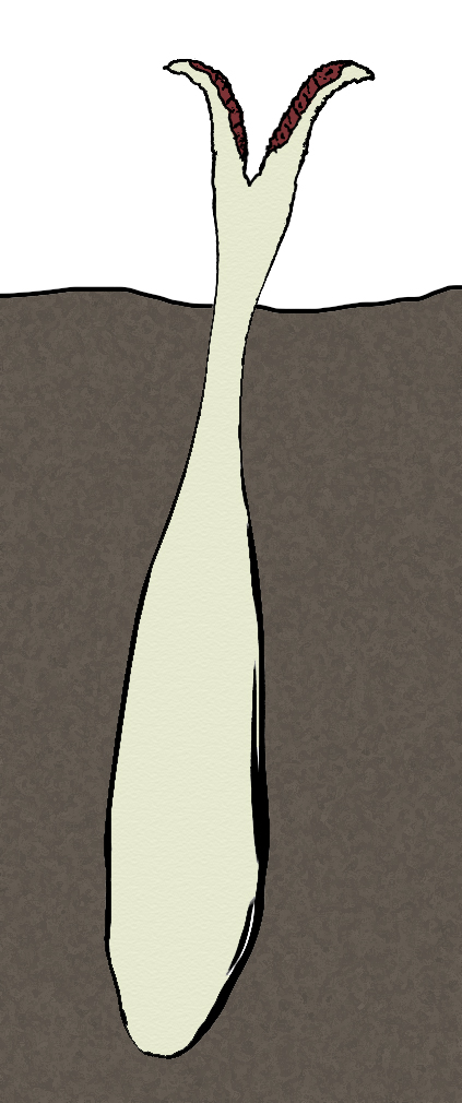
Picture by Biolog
For mushrooms great variety of shapes of sporocarps, which
frequently get rather specific and freakish shape, is typical. It is usually
characteristic for tropical mushrooms, which frequently compete to flowers in
fancifulness. But in temperate latitudes there are also rather interesting fungi
species. One of such forms is bottle fungus widespread in Eurasia.
This species belongs to the number of gasteromycetes, and its spores form in
a cavity of sporocarp. But the fungus has developed a way of spore dispersing,
using services of insects for this purpose. Bottle fungus is a saprophyte and
grows on well humidified ground in forests and among bushes. Sporocarp of bottle
fungus develops under ground; at early stages of development it has characteristic
egg-like shape and develops on the depth of up to 20 cm. Sporocarp is covered
with a double tegument: exo- and endoperidium. As sporocarp matures, endoperidium
gets characteristic bottle-like shape: lateral parts begin to grow and endoperidium
stretches hard an external tegument. At this time in sporocarp cavity is formed.
As against fungi of Geaster (earthstar) genus of human epoch, sporocarp does
not appear on the surface entirely, exoperidium does not open and does not get
the characteristic star-like shape. Further in endoperidium growth of the top
part forming “bottleneck” begins. “Bottleneck” breaks through exoperidium at
top, makes the way through ground and fallen leaves and appears on the ground
surface, towering to 12-15 cm above it.
At top of “bottleneck” sporocarp is opened via vertical crack, giving insects
an access into the internal cavity of fungus. Edges of crack bend outside, exposing
meaty-red inner side with black spots, which attracts insects by partial imitating
of meat. Internal walls of sporocarp are velvety from a friable layer of myceliums
with spores. During the time of spore-bearing sporocarp emits pleasant flowery-wine
smell. Insects fly to this smell, creep along the “bottleneck” inside the sporocarp
and eat a part of mycelia strings. Flying away from a fungus, they bear spores
on their bodies and carry them in vicinities. In shape of sporocarp of bottle
fungus and its way of attraction of insects there is a case of convergence with
plants of aroid family, at which covering leaf at an inflorescence (spathe)
forms a trap for insects of the similar shape.
After spore-bearing sporocarp of bottle fungus slowly dies off. In dry summer
and an autumn the rests of sporocarp do not decompose, but dry up into parchment-like
mass and keep till the next spring.
Antidote
bracket fungus (Fomitopsis detoxicator)
Order: Polyporales (Polyporales)
Family: Fomitopsidaceae (Fomitopsidaceae)
Habitat: Japan Islands, parasite of “death tree”.

Picture by Biolog
Japan Islands harbor one of the largest poisonous plants of Neocene epoch
– so-called “death tree”,
an arboraceous plant of Anacardiaceae family. Its toxins are so poisonous, that
it is unsafe even simply to stand in thickets of this tree. Few animals and
plants are capable to endure the neighbourhood of this species. Nevertheless,
a poisonous tree has symbiotes, and some species of animals feed on its foliage
or seeds, accumulating toxins of tree in their bodies for protection against
enemies. But the protection of “death tree” itself is not absolute, and it has
its own enemies.
On trunks of “death tree” large tinder fungi species settles – antidote bracket
fungus. It is one of few parasites on this poisonous tree. The secret of its
survival is consists in the presence of special enzymes – the powerful weapon
in chemical war plants lead against each other. This fungus is able to decompose
poisonous components of plant sap to harmless substances, and begins its expansion
on the “neutralized” areas.
The significant part of a body of fungus is hidden under bark and in the top
layer of wood of “death tree” as mycelium – richly expanding and clogging tracheae
of tree. From outside on tree bark sporocarps of this fungus are visible – perennial
(existing up to 4-5 years) ones, slightly lignificating in the centre, but soft
and spongy on edges. Diameter of mature sporocarp reaches 40 cm. The top side
is colored rusty-brown passing to edges to dark yellow. Spongy spore-bearing
layer on the bottom side of sporocarp is white. The top side of sporocarp is
flat or slightly convex in the middle, edges are slightly wavy.
Sporocarps develop on host tree trunk in small groups, young sporocarps grow
above mature ones. Usually sporocarps of antidote bracket fungus develop at
height of 3-4 meters above the ground.
This fungus removes a part of poison with the drops of liquid formed along the
edge of sporocarp. The liquid contains very small amount of poison of “death
tree”, and fungus adds to it the substances attracting insects. Due to such
trick on sporocarps of fungus beetles and flies gather, who get drunk, having
tasted a “cocktail” produced by fungus. They lose ability to fly for some time
and simply creep on the bottom side of fungus, gathering on their legs and the
bottom side of body spores, which they carry then across the forest. Spores
sprout on the damaged bark of “death tree”, and mycelium at first develops on
drops of sap of tree, penetrating them with its strings. In due course of development
mycelium penetrates deeper into the wood.
Places on trunk, where mycelium of antidote bracket fungus develops, differ
in smaller contents of poison – it is a consequence of activity of mycelium
of this fungus. It enables other fungi and animals to settle on “death tree”
and to use it as a source of food. Antidote bracket fungus destroys wood only
a little, because it is a parasite and in its development depends on well-being
of host tree. Tree infected by this fungus in great degree gradually slows its
growth rate and is exposed to an attack of wood-boring insects and also of wood-destructing
fungi, which cause oappearing of dying off sites of wood, and further the death
of a whole tree.
The idea about existence of this fungus was proposed by Anton, the forum member.
Abscess
fungus, tree sore (Xyloulcus corrosivus)
Order: Polyporales (Polyporales)
Family: Fomitopsidaceae (Fomitopsidaceae)
Habitat: forests of Europe (Western Europe, foothills of the Alpes, Fourseas
coasts) and Northern Africa (Atlas Mountains).
In Neocene in all natural communities fungi still occupy niches of decomposers
and parasites of animals and plants. In due course of evolution they began to
decompose effectively wood of both dead and live trees. One of the parasitic
fungi playing an important role in wood decomposition is an original representative
of tinder fungi which is named as tree sore, abscess fungus for original appearance
of its sporocarps. This fungus descends from sulphur polypore (Laetiporus sulphureus)
known in human epoch.
Like its ancestor, abscess fungus is a parasite of deciduous trees; it never
lives on coniferous trees. This fungus is easy to recognize due to its characteristic
appearance. Its sporocarps appear on various places of trunks of infected trees
– from roots up to inner part of crone. Their presence is easy to find out:
from such sites the bark peels, and fungus forms on the damaged wood dense mycelia
“carpet” of color from yellowish-white up to citreous and bright rusty. Under
the layer of mycelium an active process of destruction of wood takes place,
in which not only fungus takes part, but also numerous insect larvae. Fungus
decomposes also their dung containing a significant amount of non-digested cellulose.
The damaged site of wood is literally corroding – from year to year, it becomes
deeper and more extensive. Tree trunk loses durability and falls, especially
if it was infected near to roots. This fungus settles at the sites of damaged
wood not covered with bark, and further spreads to the whole tree trunk.
Sporocarps of this fungus are modified deeply in comparison with ancestral species.
Formation of a sporocarp begins in thickness of “carpet” of mycelium, where
a spherical formation appears. In due course of growth in it an internal cavity
appears, and on its surface the formation of hymenophor begins. The wall of
sporocarp directed down grows thinner and turns to a thin membrane. When growing,
sporocarp gets the shape similar to orange wedge. To the moment of complete
development of sporocarp, the membrane on the side directed down is broken off
and opens spongy hymenophor of white color. The top side of sporocarp is smooth
or slightly knobby, along the edge of sporocarp rags of protective membrane
hang down.
Spores of this species are frequently carried by insects. Formation of sporocarps
takes place more often in places of pupation of beetles, which at larval stage
were fed under the cover of mycelium. As a rule, in sporocarp a burrow may be
found, which is gnawed through by the beetle after the termination of its metamorphosis.
In warm areas abscess fungus grows the year round. At the north of its range,
in conditions of cool winter, peel with sporocarps sheds off every year and
grows again in spring of the next year from mycelium wintered in thickness of
wood.
Sporocarps and mycelia “carpet” of abscess fungus are edible for many kinds
of animals. They frequently become a food for rodents and others ground-dwelling
mammal, and also for various invertebrates, especially for snails and slugs.
This species grows rather fast; therefore it easily restores damage put by animals.
Thick
petal fungus (Crassipetalops sanator)
Order: Polyporales (Polyporales)
Family: Fomitopsidaceae (Fomitopsidaceae)
Habitat: Eurasia, old woods of various types in the temperate climatic zone.

Picture by Biolog
Tinder fungi have rather successfully endured human epoch,
settling in artificial tree plantings and invading wooden constructions. Only
few highly specialized forms of these fungi have become extinct after destruction
of their habitats: these were mainly the kinds infecting old trees of slowly
growing species inhabited mainly old-grown forests. Survived species of tinder
fungi have quickly restored their number and range after the ending of human
epoch, and among them the species occupied ecological niches of extinct forms
appeared alongside with kinds leading a new way of life.
One unusual representative of tinder fungi is thick petal fungus. This species
occupies extensive areas of woods of Eurasia in zone of temperate climate with
obviously expressed seasonal changes. It settles mainly on deciduous trees,
preferring old trees with wood heavily affected by insects.
Mycelium of this species destroys wood inactively and grows mainly in tunnels
bored by insects, clogging them with friable cotton wool-like congestions of
mycelium secreting sticky sweetish liquid. When a grub touches them, it usually
sticks to mycelium, which quickly envelops this grub and penetrates into its
spiracles and digestive path, killing it. Mycelium quickly absorbs nutrients
from grub body, accumulates them, and then uses for formation of sporocarp.
Because of this feature this species of fungi most successfully grows on trees
heavily affected by insects, and can reduce appreciably the number of wood pests.
Thick petal fungus develops rather slowly, and only at the end of summer it
forms on tree trunks smann annual sporocarps growing one by one. Sporocarp of
this fungus has unusual and easily recognizeable shape – it is very flattened,
of gristly consistence and semicircular outlines with deeply dissected edge,
which forms lobes of approximately equal size, for what this fungus is named.
Sporocarp does not lignify and keeps elasticity till early winter, and then
dies off. The top side of sporocarp is smooth, beige-white in color, gradually
passing to brown at edges. Hymenophor is spongy, of white color, plentifully
producing yellow spores.
Spores are sticky, dispersing by means of wood-boring insects. They stick to
armours of beetles during their feeding on fungus, keeping viability after passing
via intestines of insect. By means of insects they get in wood of the necessary
quality, where the new mycelium develops.
Yi
qiu mushroom (Ganoderma formicarium)
Order: Polyporales (Polyporales)
Family: Ganodermataceae (Ganodermataceae)
Habitat: tropical forests of Philippine Archipelago.
During a history of the Earth mushrooms many times entered symbiosis with various
kinds of animals, mainly with insects. This union, frequently existing during
millions years, resulted in development of close interrelation between their
species. Fungi cultivated by leaf-cutting ants are those, for example. In forests
of Philippines in Neocene epoch one species of tinder fungi has entered symbiotic
relations with one local ants
species.
This species, yi qiu mushroom, provides symbiote ants with habitation, receiving
in exchange protection from mycophagous insects and an additional feeding due
to waste formed in insect colonies. This fungus has an appearance typical for
tinder fungi and infects the weakened and old trees, preferring fast-growing
species with soft wood. Because of it, it is possible to meet this fungus more
often in places where the restoration of the damaged forest cover has entered
a finishing phase, and the trees forming a secondary forest are gradually superseded
by slowly growing species with hard wood.
The body of this species is present as mycelium stretching under a bark of infected
tree on its trunk and large branches. Sporocarps form mainly in crone of tree;
top side of it is flat, and edges are slightly raised. The bottom side of sporocarp
is inflated, hymenophor is spongy. In fact, sporocarp has almost entirely a
spongy structure suitable for life of symbiotic ants, and only from above it
is covered with dense leathery tegument. Top side of sporocarp is colored rusty-brown
shade with darker concentric lines marking the periods of temporary slowing
of growth; bottom side has ash-grey color. In good conditions diameter of sporocarp
reaches 50 cm; it accrues from edges every year, existing up to 6-8 years. Sporocarps
form on tree trunk in groups: younger ones grow from below, under old ones,
forming “racemes” up to 2-3 meters long. Usually all sporocarps are inhabited
by ants forming one or several closely related families.
Old sporocarp gradually destroys wood in an adjoining part of tree trunk, forming
the cavities lined with mycelium. In such a way fungus gets feeding from organic
remains gathered by ants: insects drag waste from nest into these cavities.
Fungus has passed to dispersing of its spores with the help of symbiotes. Spore-bearing
areas are formed on the surface of hymenophor, but only in restricted places.
The part of spores is taken outside during the ventilation of nest by ants,
but much more often spores are carried by winged breeding individuals during
a nuptial flight. Usually winged individuals of symbiote ants infect a tree
when feed on sap flowing out from damaged trunk. Also the tree appears infected,
when impregnated ant female founds a colony in a crack under tree bark. When
the first sporocarp appears, a colony of insects begins more intensive expanding.
Attracting ants, mushroom secrets a small amount of sweetish sap in thickness
of sporocarp.
The idea about existence of the present species of fungi was proposed by Morgot, the forum member.